CT scan results at Tam Anh Cau Giay General Clinic suggested a foreign body on the right esophageal wall and localized pneumothorax in the corresponding area.
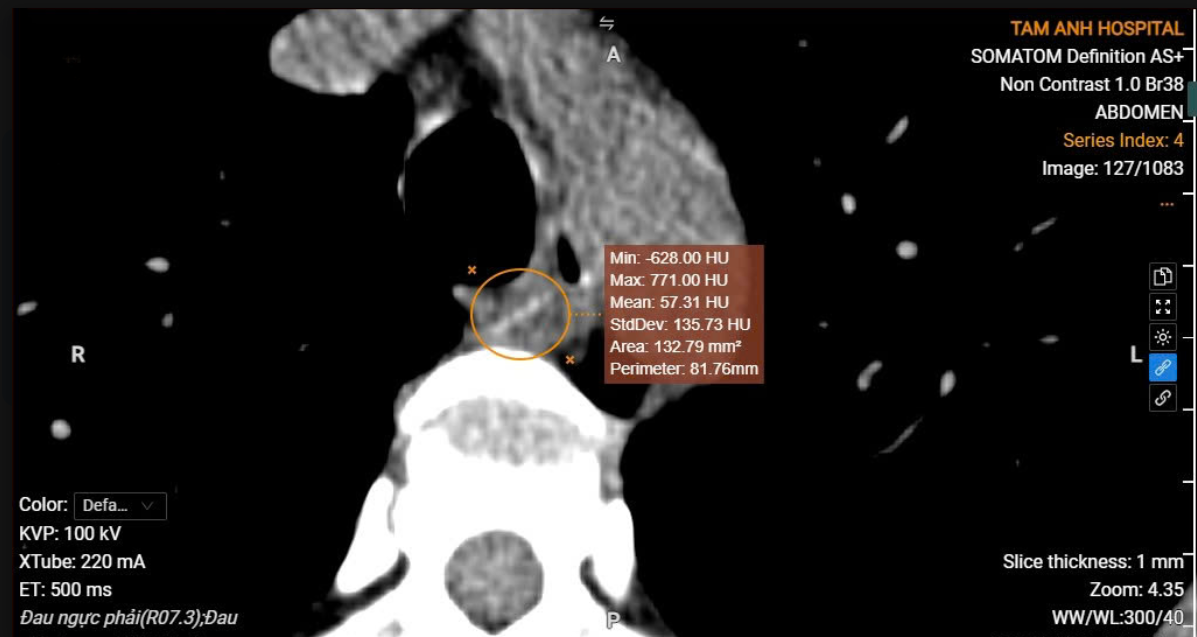 |
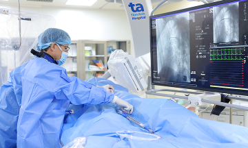
Image of fish bone on CT scan, easily overlooked if not carefully observed. Photo: Tam Anh Hospital. |
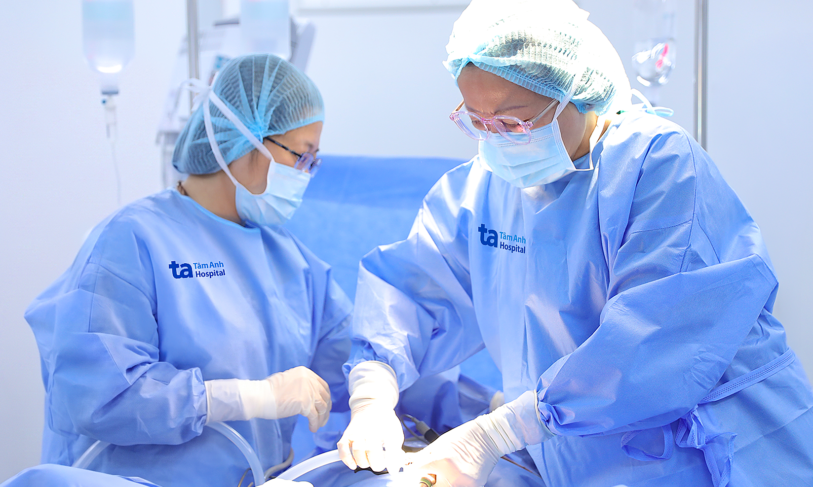
Dr. Dao Phuong Thuy, from the Department of Respiratory, ordered an esophago-gastroscopy. This procedure confirmed that a fish bone fragment had pierced the esophageal wall, damaging the pleura and causing pneumothorax.
The doctor successfully removed the fish bone. During the procedure, unusual lesions were also discovered in the stomach body. Biopsy samples confirmed gastritis and helicobacter pylori (HP) infection. Mr. Tan was closely monitored for infection risk and potential complications related to the esophageal injury, and received treatment for gastritis.
 |
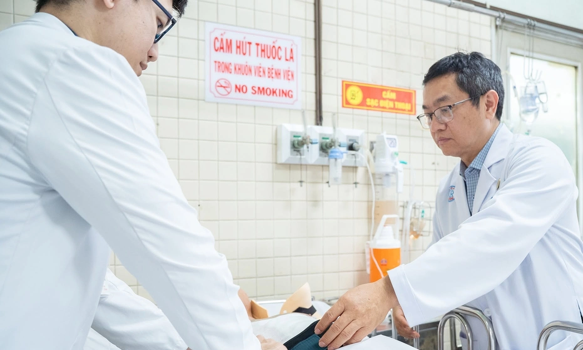
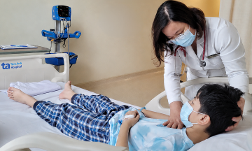
The doctor re-examines the patient before discharge. Photo: Tam Anh Hospital. |
Gastrointestinal foreign bodies, especially fish bones, are common in young children and elderly individuals. Fortunately, most cases are detected early, preventing complications.
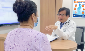
Dr. Thuy advises anyone experiencing a sensation of something stuck while swallowing, neck pain, chest pain, or prolonged discomfort after eating or drinking to seek early medical attention. She cautions against using folk remedies, such as trying to swallow rice, vegetables, or other foods to dislodge a foreign body. Such actions can embed the foreign body deeper, increasing the risk of esophageal perforation and other dangerous complications. If a foreign body is suspected, patients should go to a medical facility for timely treatment.
Khue Lam
| Readers can send questions about respiratory diseases here for doctors to answer. |