Ms. Trao reported that after her cataract surgery, her left eye did not improve. She had almost entirely lost her sight, only able to distinguish between light and dark.
An examination at the High-Tech Eye Center, Tam Anh General Hospital Hanoi, revealed the artificial lens in Ms. Trao's left eye had dislodged into the vitreous humor, the space between the lens and the retina. This was accompanied by a dense intraocular hemorrhage, a mix of old and new blood, and retinal detachment. Doctor Bui Viet Hung, Head of the Vitreoretinal Department, explained that this condition obscured the intraocular environment, making it difficult to observe internal structures, particularly the retina.
 |
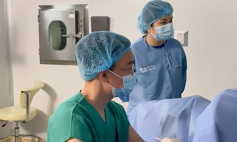
Doctor Hung examines Ms. Trao's eye before surgery. *Photo: Tam Anh General Hospital*
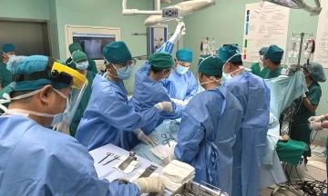
The medical team opted for a two-stage treatment plan to restore Ms. Trao's vision. During the first stage, the dense intraocular hemorrhage severely obstructed vision, rendering conventional observation tools like surgical microscopes ineffective. Consequently, the team employed an intraocular camera to directly visualize the vitreous humor, pinpoint the dislodged lens, and identify associated damage.
Ms. Trao underwent a vitrectomy to remove the blood, address retinal damage, and abnormal tissues. Concurrently, intraocular silicone oil was injected to reattach the retina and ensure its stability. Following this initial intervention, her retina had not completely lost function, allowing doctors to continue monitoring and progress to the subsequent treatment stage.
Once the retina was stable and properly reattached, Ms. Trao proceeded to the second stage of treatment. This involved removing the silicone oil and implanting an artificial lens. Doctor Hung explained that performing the lens implantation in a later stage helps ensure a stable intraocular environment. It also allows for a precise assessment of the retina's functional recovery, which is a crucial factor in predicting the patient's visual prognosis.
Following the two surgeries, Ms. Trao's left eye vision significantly improved, enabling her to recognize images. Post-operative examinations confirmed that the retina had returned to its correct position within the eye, intraocular structures were re-established, and the internal eye environment gradually stabilized. This positive outcome indicates that the retina retained functional cells, capable of receiving and transmitting light signals after treatment.
Doctor Hung emphasized that when an artificial lens dislodges into the vitreous humor, causing retinal damage, correctly addressing the root cause and preserving the retina are critical factors for restoring vision. He noted that despite the potentially lengthy treatment, timely intervention can significantly improve both vision and overall quality of life.
After cataract surgery, patients require regular follow-up appointments. Should they experience increased blurred vision, rapid vision loss, or any visual abnormalities, they should seek prompt medical examination for timely intervention.
Thu Giang
| Readers can send questions about ophthalmological diseases here for doctors to answer. |