Looking at her healthy 4-month-old son, a 30-year-old mother is filled with happiness. She still vividly remembers the moment her 28-week-old fetus was between life and death, and the journey from despair to miracle that brought her to this joyful day.
During her first pregnancy, she went to the University of Medicine and Pharmacy Hospital in Ho Chi Minh City at 28 weeks experiencing shortness of breath and fatigue. Through a fetal echocardiogram, Associate Professor Doctor Le Minh Khoi, Head of the Cardiovascular Imaging Unit, discovered a critical situation. The fetus had pericardial effusion, severe heart failure, and a giant placental tumor. This tumor, by increasing blood vessel growth, caused increased blood flow to the fetal heart, leading to circulatory overload and heart failure.
"This was a dangerous clinical situation that put the fetus's life in jeopardy," recalled Associate Professor Khoi.
At only 28 weeks, the fetus was premature, presenting significant risks if delivered early. Doctor Tran Nhat Thang, Head of the Obstetrics and Gynecology Department, explained that premature birth at this stage could lead to serious complications such as neonatal infections, brain hemorrhages, and necrotizing enterocolitis. Monitoring the fetus daily, the doctors realized they couldn't delay intervention.
A multidisciplinary team of doctors convened and agreed to perform an embolization of the blood vessels feeding the placental tumor. The goal was to relieve pressure on the fetal heart and extend the pregnancy by at least 4 weeks. According to Associate Professor Doctor Vo Tan Duc, Head of the Diagnostic Imaging Department, endovascular interventions are typically performed under X-ray guidance. However, to protect the fetus from radiation exposure, this entire procedure had to be performed under ultrasound guidance.
"Inserting a needle through the abdominal wall into the pregnant woman's uterus to accurately locate and access the tumor's main blood vessels requires high precision, coordinated teamwork, and extensive experience," the doctor explained.
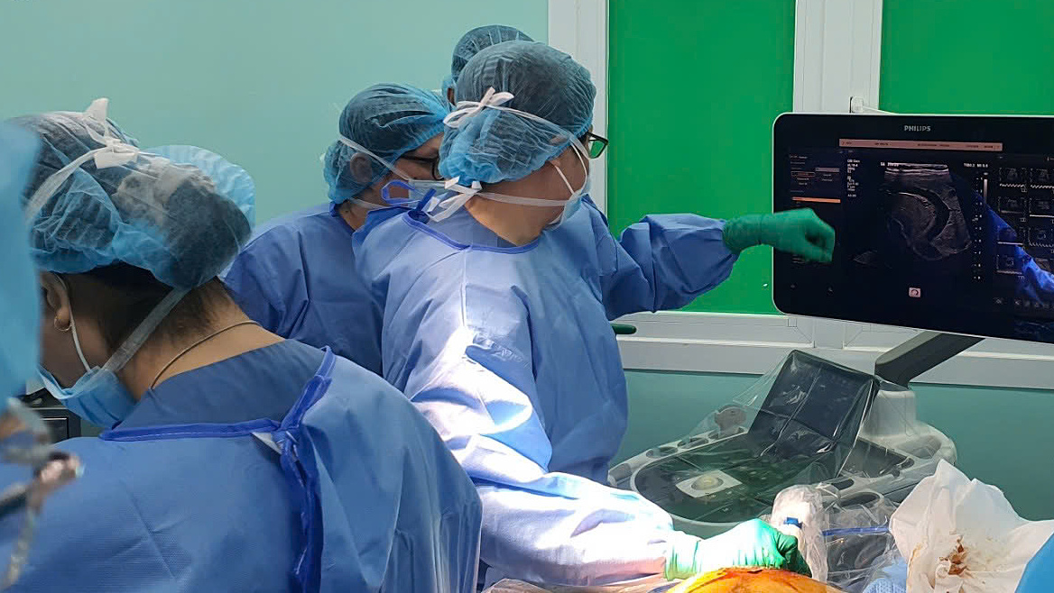 |
Medical staff performing the intervention on the pregnant woman. Photo: Hospital provided |
Initially, the team used coils (small metal springs) to embolize the tumor, but the blood flow was too strong to be completely blocked. The doctors made a pivotal decision to use bio-glue to fill the gaps around the coils and blood vessels. This procedure required near-absolute precision, as if the bio-glue entered the fetal heart or brain through the bloodstream, it could block critical vessels, potentially leading to death.
The results exceeded expectations. After the intervention, blood flow to the fetal heart decreased rapidly, the tumor shrunk, and the fetal heart recovered. The pregnancy continued not only to the initially targeted 32 weeks but extended to 39.5 weeks, resulting in a safe delivery without any emergency interventions.
The young mother said that when informed about the fetus's critical condition, despite the doctors' thorough explanations, she couldn't hide her worry and fear. It was the first time this technique had been applied and carried potential risks. Telling herself, "If I don't try, I might regret it later," she placed her trust in the medical team, giving her child a chance at life.
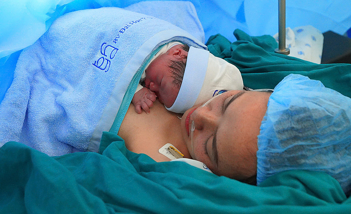 |
The newborn baby having skin-to-skin contact with the mother. Photo: Hospital provided |
Associate Professor Doctor Nguyen Hoang Bac, Director of the University of Medicine and Pharmacy Hospital in Ho Chi Minh City, stated that this was not only a professional success but also a testament to the solidarity, courage, and responsibility of the medical staff. "We didn't choose the easy path, we chose the right path, contributing to the development of fetal medicine in Vietnam," he said.
Le Phuong