Thanh, an American exchange student home in Vietnam for summer break, began experiencing frequent, severe headaches accompanied by dizziness, blurred vision, and loss of balance. Attributing his symptoms to the change in weather and jet lag, Thanh initially delayed seeking medical attention. However, when his symptoms worsened, he went to Tam Anh General Hospital in Ho Chi Minh City for an examination. A 3 Tesla MRI scan of his brain revealed a large, 4.6 cm tumor in the vermis of the cerebellum. The tumor was compressing the 4th ventricle and cerebral aqueduct, causing dilation of the lateral and 3rd ventricles, periventricular edema, and tonsillar herniation through the foramen magnum.
On 26/8, Dr. Mai Hoang Vu, from the Department of Neurosurgery - Spine at the Neuroscience Center, explained that Thanh's secondary headaches were likely related to the brain tumor. After consultation, doctors suspected it was a Medulloblastoma - a highly malignant type of embryonal neuroepithelial tumor found in the cerebellum. This type of tumor is particularly dangerous due to its rapid growth and tendency to spread through the cerebrospinal fluid. It is more common in children and adolescents.
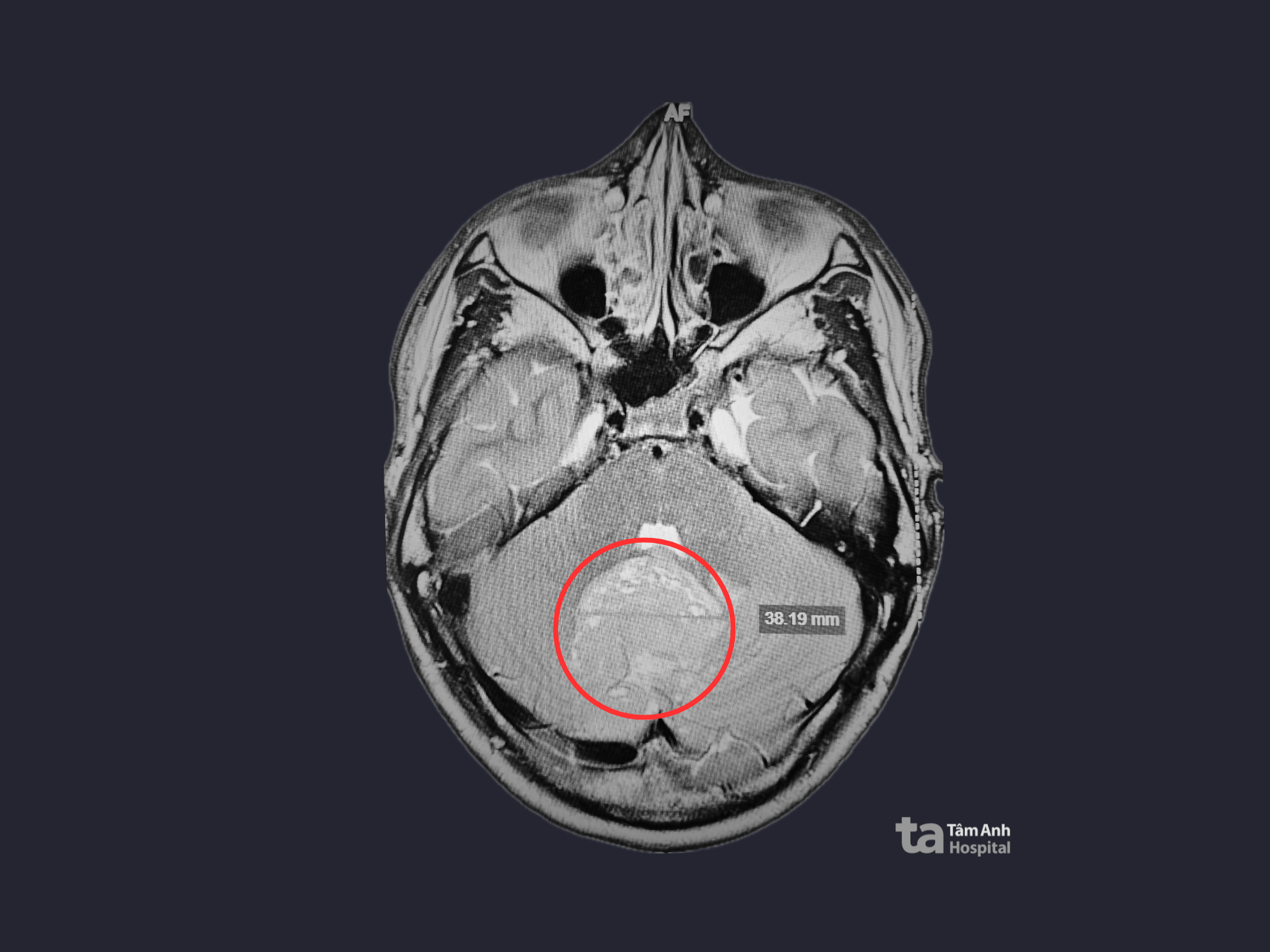 |
A 3 Tesla MRI scan revealed the tumor located in Thanh's cerebellum. Photo: Tam Anh General Hospital |
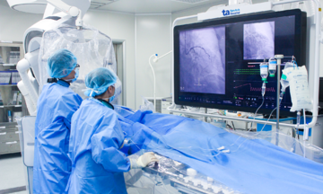
Surgery was recommended to remove as much of the tumor as possible, alleviate the cerebrospinal fluid blockage, and obtain a tissue sample for biopsy to determine the tumor's exact nature and guide further treatment. Dr. Vu anticipated a complex surgery due to the tumor's infiltrative nature into the surrounding healthy brain tissue, making it difficult to distinguish between the tumor and normal tissue. The surgical team decided to use a technique involving Fluorescein Sodium, a fluorescent dye, to guide the operation. This organic dye glows under green light at wavelengths between 540 and 690 nm.
After intravenous injection, the dye accumulates in areas where the blood-brain barrier is compromised, clearly highlighting the tumor's infiltration into the surrounding brain tissue. Using the AI Kinevo 900 surgical microscope equipped with fluorescence imaging and specialized filters, the entire boundary of the tumor became distinctly visible, separate from the healthy brain tissue, allowing the surgeon to precisely remove the diseased tissue.
The nearly three-hour surgery successfully removed almost the entire tumor, relieving the pressure on the ventricles. The biopsy confirmed the diagnosis of Medulloblastoma, SHH subgroup with wild-type TP53.
Post-surgery, Thanh was alert and responsive, with stable neurological signs. His headaches significantly subsided, the nausea and dizziness disappeared, and he began walking with the assistance of a physical therapist. He is currently under observation as doctors develop a complementary radiation and chemotherapy regimen to prevent recurrence.
 |
Surgeons removing Thanh's tumor. Photo: Tam Anh General Hospital |
Dr. Vu explained that tumors originating in the 4th ventricle tend to compress the brainstem, often initially presenting with morning headaches, nausea, blurred or double vision, unsteady gait, and even coma. He advised that individuals experiencing persistent early morning headaches accompanied by vomiting, dizziness, or balance problems should consult a neurologist without delay.
Phuong Thy
*The patient's name has been changed
| Readers can submit questions about neurological conditions here for doctors to answer. |