Nghia experienced a sudden, severe headache, collapsed, and developed left-sided weakness and slurred speech. His family rushed him to a local hospital. A CT scan revealed a right-hemisphere brain hemorrhage, prompting his transfer to Tam Anh General Hospital in TP HCM for intervention. Upon arrival, Nghia's blood pressure was high (153/94 mmHg), his left arm strength was 2/5, his left leg strength was 3/5, and his consciousness was declining. Imaging showed a roughly 6 cm hematoma in his right brain causing cerebral edema, compressing the right lateral ventricle, and shifting the midline 4 mm to the left.
On 23/9, Dr. Mai Hoang Vu, from the Neurosurgery Department of the Neuroscience Center, reported Nghia's critical condition. He needed urgent surgery to remove the hematoma, relieve the pressure, halt the toxic inflammatory cycle causing cerebral edema and further damage, and preserve as much healthy tissue as possible.
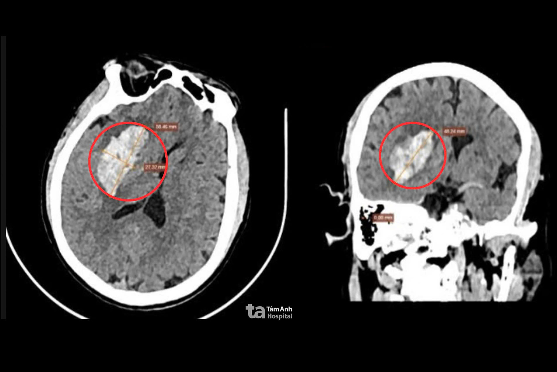 |
A CT scan revealed a large hemorrhage in Nghia's brain. Photo: Tam Anh General Hospital |
A CT scan revealed a large hemorrhage in Nghia's brain. Photo: Tam Anh General Hospital
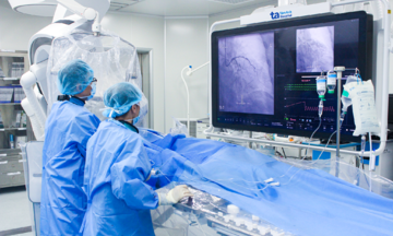
After consultation, Nghia was scheduled for minimally invasive surgery (MIS) assisted by artificial intelligence (AI). A small incision was made in his scalp, and a high-speed drill created a small, roughly 2 cm "keyhole" opening in his skull. The specialized BrainPath device was inserted deep into the brain, selectively following natural grooves. Surgeons gently separated brain tissue to create a safe corridor to the hematoma without damaging healthy tissue.
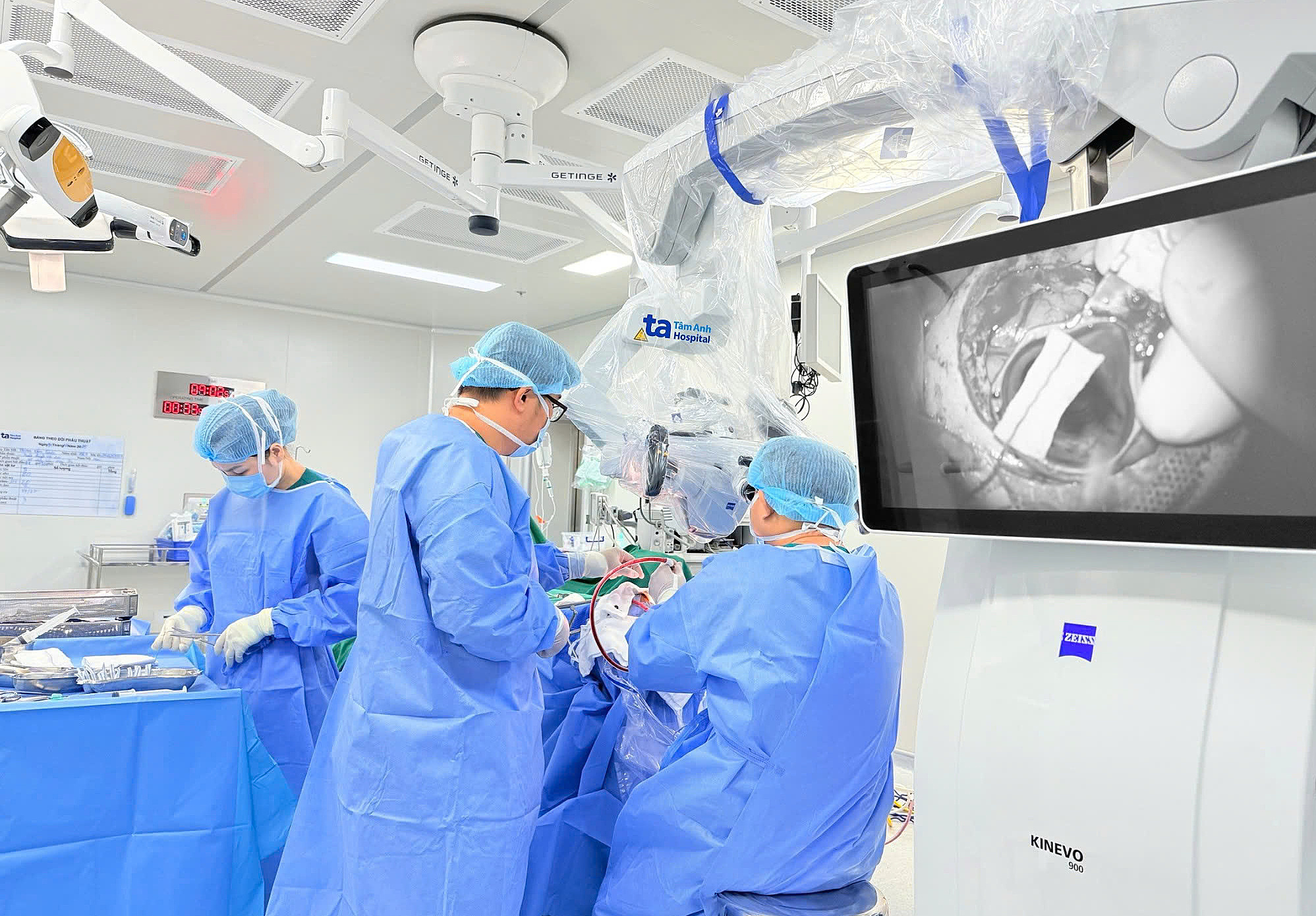 |
Surgeons removing Nghia's hematoma using minimally invasive techniques. Photo: Tam Anh General Hospital |
Surgeons removing Nghia's hematoma using minimally invasive techniques. Photo: Tam Anh General Hospital
Using the AI-powered Zeiss Kinevo 900 microscope with 3D imaging, the surgical team precisely located the hematoma, visualized even small blood vessels, and carefully removed the clot without causing further trauma. FloSeal hemostatic material was used to minimize the risk of re-bleeding.
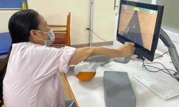
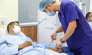
Post-surgery, Nghia was conscious and didn't require intensive care. His incision was clean and dry. Three days later, he showed good recovery, improved consciousness, and regained leg movement. A follow-up CT scan confirmed complete hematoma removal, with only slight ventricular compression and a normalized midline. Nghia's vital signs were closely monitored to prevent re-bleeding. After managing his underlying conditions and stroke risk factors, he was discharged and continued rehabilitation with a personalized exercise plan.
Doctors advise individuals with underlying conditions like hypertension, diabetes, and dyslipidemia to have regular checkups, adhere to treatment plans, and maintain a healthy lifestyle. If experiencing symptoms such as weakness, difficulty speaking, facial drooping, or severe headaches, seek immediate medical attention at a stroke-specialized facility.
Phuong Thy
*The patient's name has been changed.
| Readers can submit questions about neurological conditions here for doctors to answer. |