On 4/9, Doctor Truong Long Vy, head of neurosurgery at Thu Duc General Hospital, reported that a CT and MRI scan revealed a 3.5x5 cm meningioma in the patient's left temporal lobe. The patient had several underlying conditions, including diabetes, hypertension, and coronary artery disease, which were not well-controlled. A multidisciplinary team of doctors coordinated preoperative treatment, including discontinuing anticoagulants 5 days prior to the procedure.
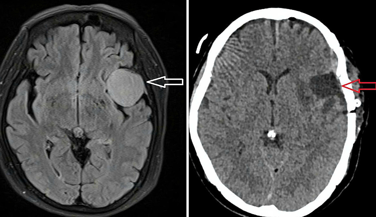 |
The patient's brain tumor before (left) and after (right) surgery. Photo: Hospital provided |
The patient's brain tumor before (left) and after (right) surgery. Photo: Hospital provided
The surgical team utilized specialized equipment for cranial microsurgery, including a Mayo head frame, a surgical microscope, and a high-speed drill. The lengthy operation presented difficulties due to the thick dura mater adhering to the skull. The surgeons successfully removed the entire tumor without damaging surrounding structures.
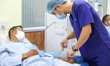
Post-operatively, the patient received specialized care in the intensive care unit, under deep sedation for 48 hours to protect neurological function. She has since recovered, is alert, communicates well, and continues to be monitored at the hospital.
The patient's daughter admitted to initially attributing her mother's headaches to old age, never suspecting a large tumor. "Fortunately, the disease was detected and treated promptly, allowing my mother to recover," she said.
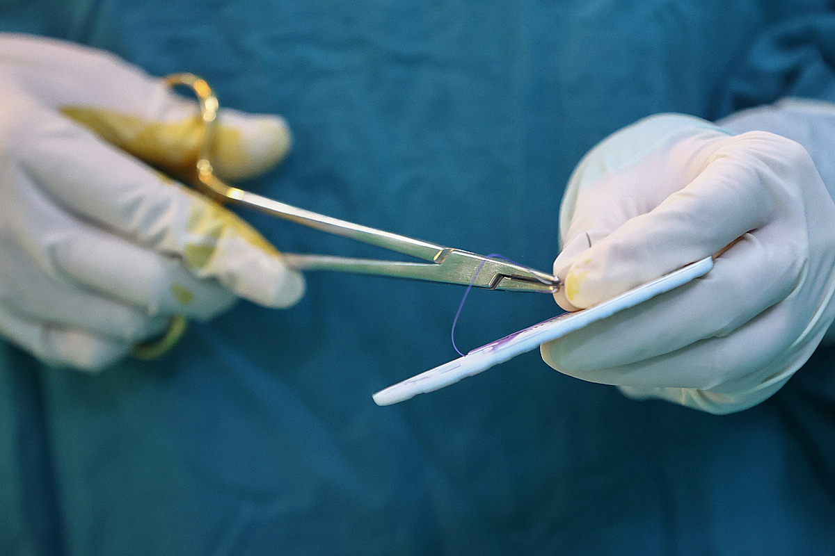 |
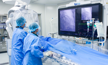
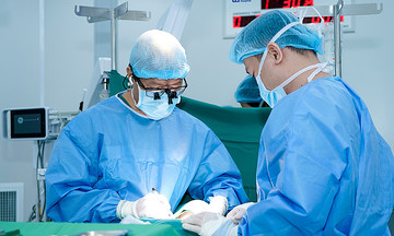
Doctors in the operating room during the patient's surgery. Photo: Quynh Tran |
Doctors in the operating room during the patient's surgery. Photo: Quynh Tran
According to Doctor Vy, persistent headaches are not always indicative of a brain tumor. However, if accompanied by neurological symptoms such as visual disturbances, limb weakness, or seizures, prompt medical attention is crucial. Early detection facilitates more effective treatment and prevents tumor growth, which can pose serious risks.
Le Phuong