Mr. Manh suffered a stroke 10 years ago and is currently being treated for hypertension and lipid metabolism disorder. Recently experiencing unusual symptoms, he visited the Tam Anh General Clinic in District 7. MRI and CT scans of his coronary arteries revealed damage to the left frontal lobe (the site of a new stroke), and chronic blockage of the left internal carotid artery (a major blood vessel in the neck that supplies oxygen-rich blood to the front and middle parts of the brain). Two arteries supplying blood to the heart, primarily the left ventricle, were significantly narrowed: the left anterior descending artery and the left main coronary artery.
He was transferred to Tam Anh General Hospital in TP HCM. Professor Vo Thanh Nhan, Director of the Interventional Cardiology Center, reported that Mr. Manh had suffered a moderately sized stroke. His left internal carotid artery was completely blocked, but his collateral circulation (the network of smaller blood vessels activated to supply blood to the brain in place of the blocked artery) was good. He did not exhibit symptoms such as unilateral weakness, severe headache, language or vision disturbances. Doctors prescribed optimal medical treatment to manage the stroke.
The severe narrowing of the left anterior descending and left main coronary arteries caused ischemia in a large area of the heart, placing Mr. Manh at high risk of death from an acute myocardial infarction (heart attack). Professor Nhan assessed that immediate coronary stenting, which requires dual antiplatelet therapy and anticoagulants to prevent blood clots, could worsen the unstable acute stroke. The doctors decided to closely monitor his condition, preventing complications such as cerebral hemorrhage, heart attack, arrhythmia, and heart failure. One month later, after the stroke was fully controlled, the team decided to perform coronary stenting.
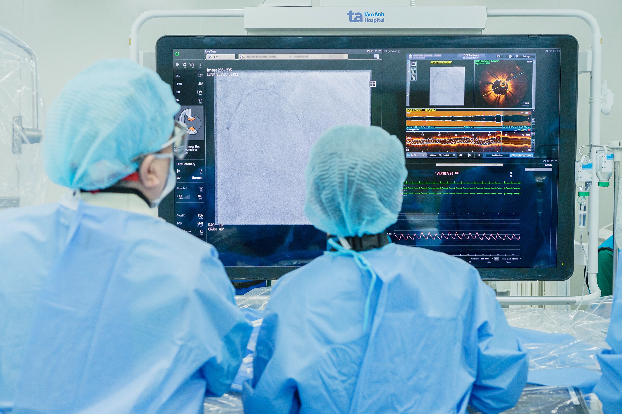
Dr. Nguyen Van Duong of the Interventional Cardiology Center used optical coherence tomography (OCT) to precisely determine the location, length, and diameter of the narrowed coronary artery segments to select the appropriate stent size for Mr. Manh. The team placed two stents in the blocked sections of the left anterior descending and left main coronary arteries. Post-procedure, both stents were fully expanded and flush against the vessel walls, with no dissection at either end.
Three days later, Mr. Manh was discharged in stable condition and scheduled for follow-up appointments. The current treatment goals are to prevent blood clots within the stents and prevent stroke recurrence. Mr. Manh is adhering to his medication regimen, following a low-salt diet, limiting starch, sugar, skin, fat, liver, and animal organs, and exercising regularly.
 |
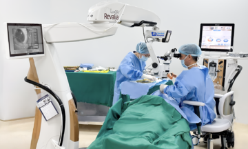
Professor Nhan (left) and his team perform stenting on a patient with the assistance of optical coherence tomography. Illustrative photo: *Tam Anh General Hospital* |
Most cases of heart attack and stroke stem from atherosclerosis. This involves the buildup of plaque – composed of cholesterol, fat, calcium, and inflammatory cells – inside artery walls. These plaques narrow the arteries, reduce their elasticity, and make them harder.
Heart attack and stroke share several risk factors, including hypertension, dyslipidemia, diabetes, and obesity. Heart attack patients have a higher risk of stroke than those without the disease. Stroke can cause serious brain damage, and cardiovascular complications include arrhythmias, cardiomyopathy, acute heart failure, and an increased risk of myocardial ischemia. Therefore, individuals with either condition should be screened for the other to enable early detection, prevention, and recurrence management.
Ngoc Chau
* The patient's name has been changed
| Readers can submit questions about cardiovascular diseases here for doctors to answer. |