Mr. Hong had undergone ureteral stone surgery twice but did not attend follow-up appointments, only seeking medical attention when his condition worsened.
A CT scan at Tam Anh General Hospital in Hanoi revealed that Mr. Hong's left kidney function had declined due to hydronephrosis. His left ureter had a stone measuring approximately 5x6 mm, and doctors suspected a stricture below the stone. Several smaller stones were also present in the lower calyx of the left kidney. While his right kidney function was normal, a few small stones were scattered in the calyces.
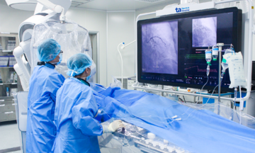
Associate Professor Doctor Tran Van Hinh, Head of the Department of Urology and Andrology, recommended retrograde ureteroscopy for Mr. Hong to both remove the stone and address the stricture, restoring urine flow from the kidney to the bladder. The patient's ureter was scarred and narrowed, making it difficult to access the stone. The surgical team used high-frequency laser to widen the ureter, allowing the endoscope to pass through.
Doctors located the stone, fragmented it with a laser, and removed all the fragments. A retrograde JJ stent was placed in the left ureter, with both ends secured in the renal pelvis and bladder to maintain urine flow and reduce the risk of re-stricture.
 |
Associate Professor Hinh examining and advising the patient. Photo: Tam Anh General Hospital |
Associate Professor Hinh examining and advising the patient. Photo: Tam Anh General Hospital
Following the procedure, Mr. Hong recovered quickly, with no further back pain, fever, or appetite issues. His kidney function also improved. He was discharged after two days in stable condition and able to resume normal activities. However, he was advised to maintain a healthy lifestyle and diet to prevent further stone formation, take prescribed medications, and attend regular check-ups.
Mai Anh
*The patient's name has been changed.