Mr. Phong's MRI revealed a carotid plexus tumor deep within the petrous bone and cavernous sinus (anatomical structures in the temporal skull base). The tumor had spread to his middle ear, compressing the internal carotid artery and cranial nerves.
"Carotid plexus tumors are rare, with only a few similar cases documented in medical literature," said Dr. Nguyen Duc Anh, Head of the Neurosurgery and Spine Department at Tam Anh General Hospital, Hanoi. He added that Mr. Phong's tumor was located in a complex area of the skull base, near major blood vessels, posing a high risk of sudden death if left untreated. The tumor's extensive growth within the petrous bone and its close proximity to the internal carotid artery (the brain's primary blood supply) made surgery particularly challenging. Any misstep during the procedure could lead to a ruptured artery, massive bleeding, nerve damage, or cerebral infarction.
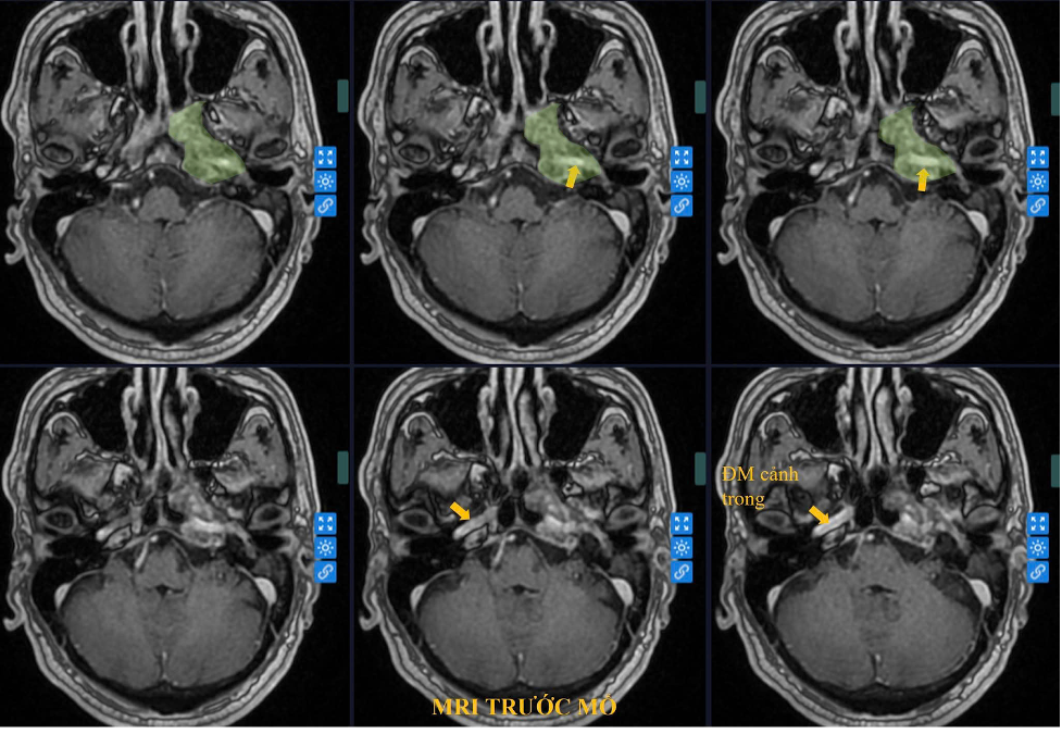 |
MRI image of the deep-seated tumor in the petrous bone. Photo: Tam Anh General Hospital |
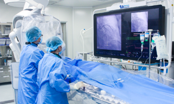
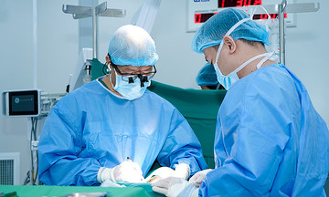
The surgical team opted for a skull base surgery approach using a surgical microscope with over 40x magnification. They also utilized the Neuro Navigation system with AI integration and intraoperative neuromonitoring (IONM), allowing for detailed pre-operative planning. The AI-integrated navigation system provided precise guidance to the tumor, differentiating it from surrounding structures and minimizing the risk of damage to healthy tissue.
Dr. Duc Anh explained that traditional surgery would have been like "navigating in the dark" due to the tumor's depth and the complex anatomical structures involved.
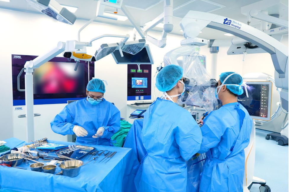 |
The surgical team using the AI-integrated Neuro Navigation system during the operation. Photo: Tam Anh General Hospital |
The team carefully drilled through the skull base layer by layer under microscopic vision, meticulously separating the tumor from the bone and the carotid artery. After 8 hours, the entire tumor was removed, and the skull base was reconstructed using artificial materials. The surgical site was then closed.
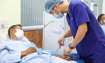
Three days post-surgery, the patient was conscious, showed no signs of nerve damage, and had normal motor function. A biopsy confirmed the tumor to be a carotid plexus tumor.
Skull base tumors, if detected late and compressing major blood vessels like the internal carotid artery, become difficult to treat. As the tumor grows, it can cause facial paralysis, vision loss, stroke, or even death. Doctors recommend that individuals experiencing persistent hearing loss, tinnitus, or deep pain in the ear-temple area consult a neurologist or ENT specialist for examination, diagnosis, and prompt treatment.
Linh Dang
*The patient's name has been changed.
| Readers can submit questions about neurological conditions here for doctors to answer. |