Mr. Vien's initial tumor measured 6 cm, and a CT scan also revealed a 1 cm nodule in the upper lobe of his right lung. At that time, Dr. Le Thi Ngoc Hang, from the Department of Thoracic and Vascular Surgery at Tam Anh General Hospital in Ho Chi Minh City, performed a minimally invasive biopsy of the lung nodule and mediastinal mass to determine a diagnosis. Results showed the lung nodule was due to a fungal infection, while the mediastinal mass was a benign thymoma. As the mass was not cancerous and not yet compressing surrounding organs, the doctor did not recommend surgery but treated the fungal lung infection for three months. Mr. Vien's health gradually recovered; the fever, cough, and chest pain subsided.
A month ago, Mr. Vien's chest pain returned. Dr. Nguyen Anh Dung, Head of the Department of Thoracic and Vascular Surgery at Tam Anh General Hospital in Ho Chi Minh City, diagnosed the tumor had increased in size to 10x8x5 cm, located near the heart and close to the aorta and vena cava. At this point, the patient's lung infection was under control, and his respiratory function was good, making him suitable for surgery to remove the mass.
Dr. Dung assessed that conventional minimally invasive surgery would be difficult due to the large size of the tumor and its proximity to the heart and major blood vessels. Open surgery would require a 10-15 cm incision and sawing through the sternum, increasing the risk of infection, blood loss, and postoperative circulatory and respiratory complications, as well as prolonging recovery time. To minimize these risks, the surgical team decided to perform a tumor resection using the Da Vinci Xi robotic surgical system. The surgery was completed quickly within an hour, with the tumor removed entirely through 4 small (8 mm) incisions.
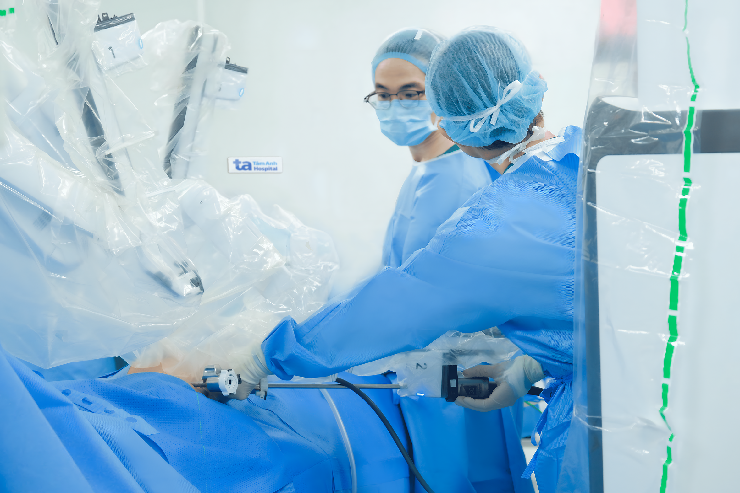 |
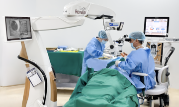
The surgical team uses robotic arms to remove the thymoma. Photo: Thanh Luan |
The surgical team uses robotic arms to remove the thymoma. Photo: Thanh Luan
Mr. Vien recovered quickly and was discharged after two days. The pathology report confirmed early-stage thymic carcinoma with no invasion. An oncologist examined Mr. Vien and recommended adjuvant chemoradiotherapy to prevent recurrence.
The thymus is located behind the sternum and plays a vital role in the development of T lymphocytes (T cells), white blood cells that fight infection. The thymus is composed of two cell types: epithelial cells and lymphocytes. Both thymomas and thymic carcinomas are thymic epithelial tumors (TETs), but thymomas are benign (slow-growing and rarely spread beyond the thymus). Thymic carcinoma is malignant, grows rapidly, and is more likely to metastasize to other parts of the body.
In the early stages of tumor formation, patients often show no clear symptoms. Some individuals remain asymptomatic until the tumor grows large and compresses surrounding structures. If symptoms suggestive of a thymoma appear, such as chest pain, coughing up blood, hoarseness, or shortness of breath, patients should seek medical attention for prompt diagnosis and treatment.
Thu Ha
*The patient's name has been changed.
| Readers can submit questions about cardiovascular diseases here for doctors to answer. |