A contrast-enhanced CT scan at Tam Anh General Hospital TP HCM revealed a tumor measuring nearly 15 cm located in the splenic flexure of the colon. This mass occupied most of the left abdominal cavity and caused colonic intussusception.
A colonoscopy identified a large tumor at the splenic flexure, nearly completely obstructing the bowel lumen, preventing the scope from passing. The tumor surface was ulcerated. Ms. Linh's abdominal cavity contained minimal fluid, with significant bowel distension and bilateral pleural effusion. Initial biopsy results suggested colon cancer.
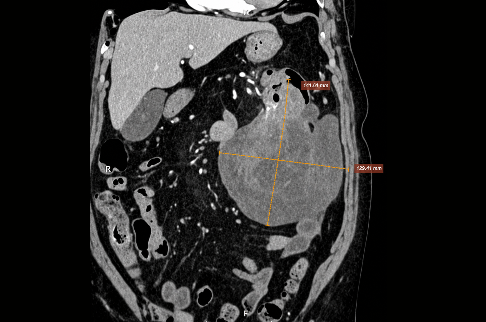 |
CT image showing a large tumor in the colon (yellow line). *Photo: Tam Anh General Hospital* |
Ms. Linh, an elderly patient, presented with a large tumor in a complex location. Dr. Nguyen Quoc Thai, Head of the Department of Gastrointestinal Surgery, Endoscopy and Gastrointestinal Endoscopic Surgery Center, assessed that surgery posed significant risks for the patient. However, untreated intussusception could constrict blood vessels supplying the bowel, leading to necrosis, perforation, and abdominal infection. The tumor nearly completely obstructed the colon lumen, posing a high risk of bowel obstruction. As the tumor grew, it risked invading vital organs such as the spleen, pancreatic tail, stomach, and diaphragm.
After consultation, doctors decided to perform open surgery to remove the entire tumor for Ms. Linh.
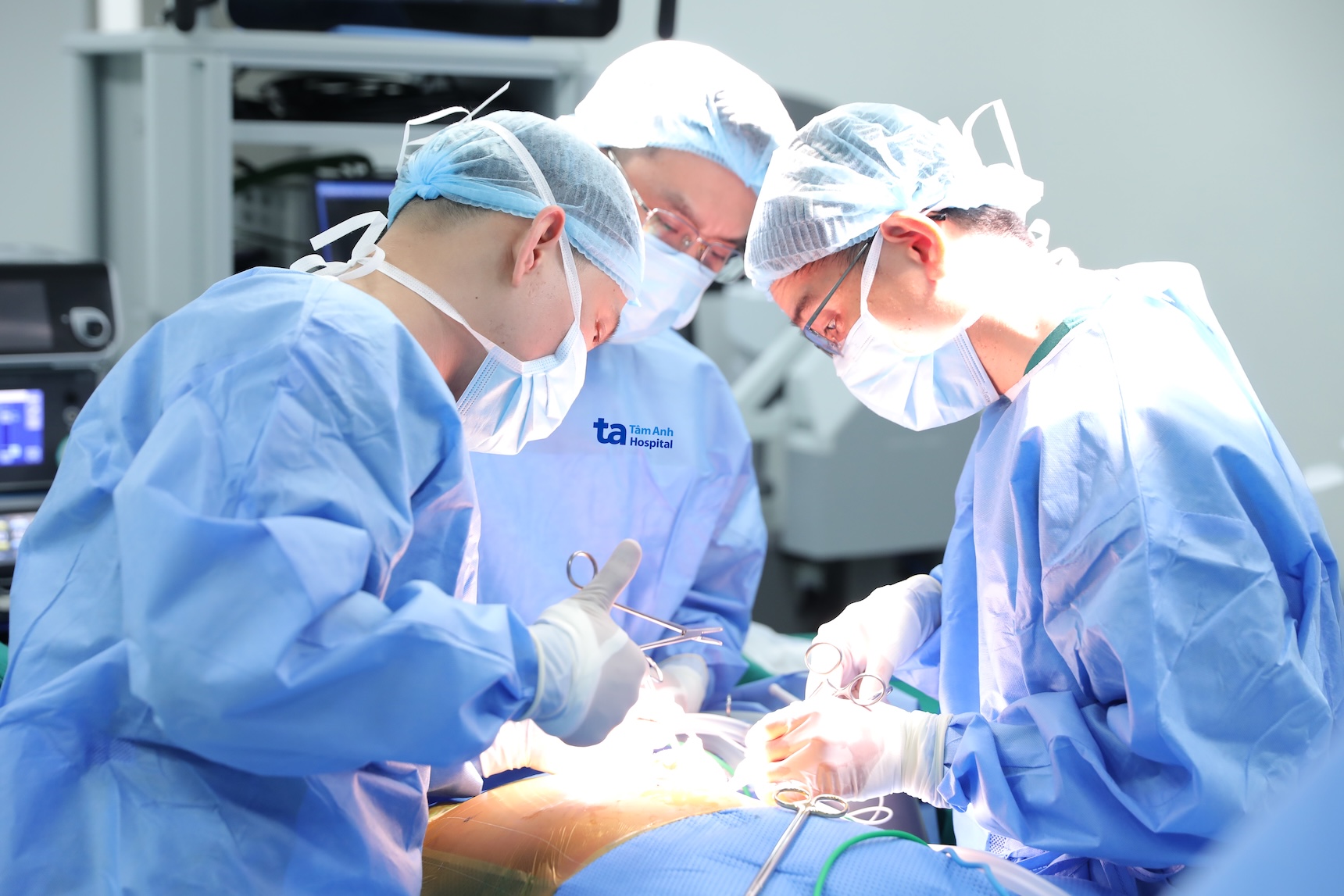 |
Dr. Thai (far right) performing colon tumor surgery for Ms. Linh. *Photo: Hospital provided* |
During surgery, the medical team observed that the tumor originated from the splenic flexure of the colon and extended to the descending colon, without invading adjacent organs. Surgeons resected the colon segment containing the tumor, performed lymph node dissection, and reconnected the two ends of the colon.
Following surgery, Ms. Linh recovered favorably, experienced no complications, was able to move gently, and consume liquid food. She was discharged after one week.
Pathology results confirmed leiomyosarcoma – a type of malignant cancer. All 7 lymph nodes removed were negative, and resection margins were clear of cancer cells. The patient did not require additional treatment.
According to Dr. Thai, colorectal leiomyosarcoma accounts for less than 0.1% of all colorectal cancer cases. Because it develops from the deep muscle layer of the intestinal wall, leiomyosarcoma often causes no clear symptoms in its early stages. Patients are usually discovered when the tumor has grown large, causing abdominal pain, changes in bowel habits, bloody stools, or a palpable abdominal mass.
Unlike common colon cancer, which typically spreads to lymph nodes first, leiomyosarcoma tends to metastasize hematogenously, most commonly to the liver and lungs. The tumor also possesses a strong capacity to invade adjacent organs and spread widely within the abdominal cavity.
Detecting the disease at a resectable stage improves prognosis and reduces the risk of recurrence or distant metastasis.
Quyen Phan
*Patient's name has been changed
| Readers can send questions about digestive diseases here for doctors to answer. |