A 3 Tesla MRI scan at Tam Anh General Hospital in Ho Chi Minh City revealed a nearly 2.5 cm tangled mass of abnormal blood vessels in the right parietal region of 34-year-old Nguyen Ngoc's brain. The feeding artery originated from a branch of the right middle cerebral artery, draining into a superficial vein.
On 25/7, Dr. Chu Tan Si, Head of Neurosurgery, diagnosed Ngoc with a complex cerebral AVM. This condition occurs when arteries and veins connect directly without capillaries, creating a tangled mass of blood vessels. This increases blood flow pressure and carries a high risk of rupture. Intervention is crucial to prevent intracranial hemorrhage, loss of consciousness, and potentially death.
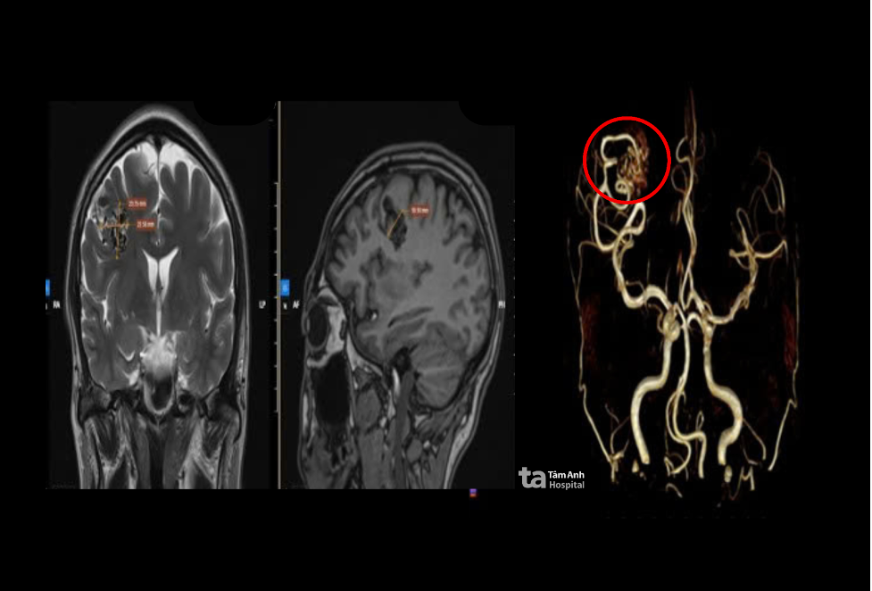 |
A 3 Tesla MRI image reveals the arteriovenous malformation in Ngoc's right parietal region. Photo: Tam Anh General Hospital |
A 3 Tesla MRI image reveals the arteriovenous malformation in Ngoc's right parietal region. Photo: Tam Anh General Hospital
Doctors decided to surgically remove the AVM. Before the procedure, Ngoc received an arterial catheter for continuous blood pressure monitoring. During surgery, an AI-integrated microscope provided magnified views of brain tissue. An AI-powered neuronavigation system pinpointed the AVM's location, feeding arteries, draining veins, and surrounding functional brain areas, enabling precise removal of the abnormal vessels. The surgical team closed the draining vein using microsurgical sutures, eliminating abnormal blood flow and minimizing damage to healthy tissue.
Ngoc recovered well, showing no signs of neurological damage, and was discharged after a week.
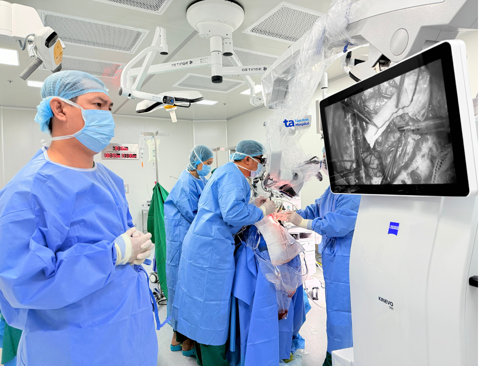 |
Doctors monitor the procedure during Ngoc's AVM removal surgery. Photo: Tam Anh General Hospital |
Doctors monitor the procedure during Ngoc's AVM removal surgery. Photo: Tam Anh General Hospital
Brain AVMs are vascular abnormalities often present from birth, frequently asymptomatic for extended periods. Larger AVMs or ruptures can cause severe headaches, seizures, hemiplegia, loss of consciousness, or even death.
According to Dr. Si, many cases are discovered only after a rupture and brain hemorrhage. This presents a high mortality risk, and survivors often experience permanent neurological damage. Advances in MRI, CT angiography, neuronavigation, and neurosurgery now allow for early detection and complete AVM removal, even before complications arise. Depending on the location and severity, treatment options include open surgery, endovascular intervention, or gamma knife radiosurgery.
Doctors recommend neurological consultation for individuals experiencing persistent headaches, fainting, seizures, or with a family history of brain AVMs for timely diagnosis and treatment.
Phuong Thy
| Readers can submit questions about neurological conditions here for doctor's responses. |