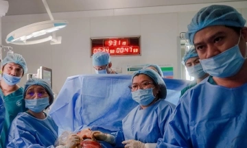
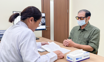
Ms. Thi's CT scan revealed an abnormal solid nodule, 1,2 cm in size, in segment S3 of her right upper lung lobe. Associate Professor Doctor Vu Huu Vinh, Director of the Thoracic - Vascular Surgery Center at Tam Anh General Hospital, Ho Chi Minh City, stated that the patient's right lung lacked a middle lobe, having only two lobes instead of the usual three. This structural abnormality is likely a congenital malformation.
"This is the first time I have encountered such a case," Associate Professor Vinh said, noting that medical literature records only a few isolated instances. He explained that some individuals have indistinct or absent fissures in the right lung, causing the middle and upper lobes to merge, but these still retain full vascular and bronchial structures. This occurs in about 11-35% of cases and differs from Ms. Thi's condition of a completely absent right middle lobe.
Associate Professor Vinh explained that when a lung lobe is absent, the remaining two lobes compensate for many years, allowing the patient to live normally without detecting the condition. However, prolonged overworking of the lungs can make these lobes more vulnerable to damage, promoting the formation of malignant tumors.
 |
CT scan showing a 1,2 cm solid nodule in segment S3 of the right lung. *Photo: Tam Anh General Hospital*
The medical team identified the greatest challenge of the surgery as the abnormal anatomical structure. The patient's lung completely lacked a middle lobe, and the vascular and bronchial systems were displaced, intertwined, and without clear separating fissures. Traditional endoscopic surgery would offer limited visibility in a confined space, making precise localization difficult and increasing the risk of incorrect dissection or damage to important vascular structures. Open surgery, while providing better visibility, would cause significant trauma, considerable blood loss, and negatively impact recovery.
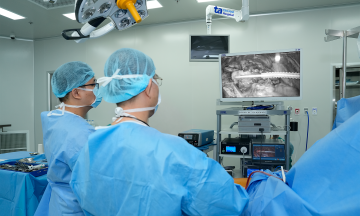
Following a consultation, the team opted for robotic-assisted endoscopic surgery using the Da Vinci Xi system. Doctors used images reconstructed from pre-operative CT scans, simultaneously evaluating them with magnified 3D camera views, to guide the robotic arms to access the lesion and collect tissue samples for biopsy. The frozen section biopsy confirmed a malignant tumor, stage T1b, with no lymph node involvement or metastasis. The team resected the S3 segment containing the tumor to eradicate the cancer and preserve healthy lung tissue, ensuring the patient's respiratory function.
 |
Associate Professor Vinh operating the robot for radical cancer surgery. *Photo: Tam Anh General Hospital*
Ms. Thi recovered quickly after surgery, with her respiratory function well-preserved. She could perform light activities and was discharged after three days. Due to early tumor detection and clear margins of malignant cells, the patient did not require additional post-operative treatment.
Early-stage lung cancer often presents no clear symptoms, leading to late diagnosis or missed detection. Associate Professor Vinh advises older individuals, current or former smokers, those working in dusty environments, or with a family history of lung cancer to undergo regular health check-ups and screenings as recommended by a doctor. Cases with congenital lung malformations or structural abnormalities also require regular monitoring for early detection of lesions and appropriate intervention.
By Bao Anh
*Patient's name has been changed.