Nguyen Tuan experienced a dull ache in his right flank for several months, which pain medication did not alleviate. Ultrasound and urinary system X-ray results at Tam Anh General Hospital Ho Chi Minh City revealed a branched coral stone, approximately 4x5 cm, that had spread to fill the shape of his renal pelvis and calyces.
Dr. Nguyen Tan Cuong, Deputy Head of the Urology Department and Head of the Urological Cancer Unit at the Center for Urology - Nephrology - Andrology, explained that coral stones are one of the most complex forms of urinary tract stones. The treatment goal is to completely remove the stones, minimize the risk of bleeding and infection, and preserve kidney function for the patient. The medical team opted for minimally invasive percutaneous nephrolithotomy (mini-PCNL) instead of open surgery. This technique provides sufficient access to fragment the entire stone with a high-power laser, limiting blood loss.
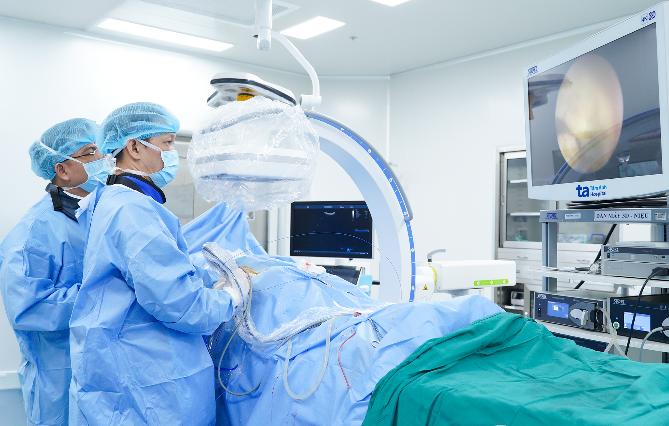 |
Dr. Cuong (right) performing minimally invasive percutaneous nephrolithotomy on Nguyen Tuan. Photo: Tam Anh General Hospital
The medical team combined 3D ultrasound and C-arm imaging to create access to the kidney. The 3D ultrasound system generates three-dimensional images, identifying the stone-containing renal calyces, guiding the approach, and recognizing blood vessel structures to avoid. Meanwhile, the C-arm system provides real-time X-ray images, allowing the team to control the needle's path, instrument placement, and depth when accessing the renal pelvis and calyces system. This combination enhances accuracy during tunnel creation, limits tissue damage, and reduces the risk of complications.
Just 90 minutes after needle puncture and creating a small tunnel, approximately 5 mm in diameter, from the skin into the kidney, the medical team fragmented the coral stone using a high-power laser and suctioned it out. Afterward, the endoscopic team meticulously inspected each renal calyx, sequentially removing any remaining stone fragments.
At the end of the procedure, doctors placed a JJ stent connecting the kidney to the bladder and a percutaneous nephrostomy tube for monitoring. Post-surgery, the patient began eating soft food early, and his urine gradually cleared from a pale pink color. The surgical wound remained dry, with minimal pain.
One day after surgery, Nguyen Tuan had his nephrostomy tube and urinary catheter removed, and he was discharged after two days. Two weeks later, he returned for a follow-up appointment to have the JJ stent removed and received advice on preventing stone recurrence.
Coral stones are often associated with chronic urinary tract infections or metabolic disorders that lead to continuous stone formation. Because they occupy nearly the entire renal pelvis and calyces system, coral stones are more challenging to remove completely in a single intervention compared to common solitary stones. If left untreated, coral stones can cause prolonged hydronephrosis, frequent recurrent infections, and even kidney dysfunction.
According to Dr. Cuong, for kidney stones larger than 20 mm, including coral stones, percutaneous nephrolithotomy is the optimal treatment method. It offers a higher stone-free rate and requires fewer supplementary interventions compared to extracorporeal shock wave lithotripsy or ureteroscopy. The minimally invasive percutaneous nephrolithotomy technique helps reduce kidney tissue damage and limits bleeding during and after surgery, which is the most concerning complication when treating coral stones with larger percutaneous tunnels.
Doctors advise patients to drink 2 to 2,5 liters of water daily, reduce salt intake, and limit animal protein to prevent disease recurrence. High-risk individuals should undergo examinations, stone composition analysis, and metabolic tests to identify causes and implement preventive measures. Patients experiencing prolonged flank pain, painful urination, or frequent urination should seek early medical attention for timely and minimally invasive diagnosis and treatment.
Ha Thanh
*Patient's name has been changed