Six months ago, Tin began experiencing a dull chest pain. A CT scan revealed a 54 mm thoracic aortic aneurysm. Although doctors recommended intervention, Tin opted for medical management and regular monitoring. Three months later, the chest pain intensified, accompanied by discomfort above his navel, nausea, and loss of appetite. A new CT scan at Tam Anh General Hospital in Ho Chi Minh City showed the existing aneurysm and an appendiceal mucocele.
Doctor Nguyen Anh Dung, Head of the Department of Cardiothoracic and Vascular Surgery, diagnosed the aneurysm extending from Tin's chest to the upper part of his kidneys. It measured 54 mm at its widest point and 30 mm at its narrowest. Dr. Dung recommended prompt intervention, either a stent graft placement or surgery, to prevent complications like rupture, thrombosis, heart failure, stroke, or sudden death. The mucocele's nature – benign or malignant – was undetermined, but it carried a high risk of infection.
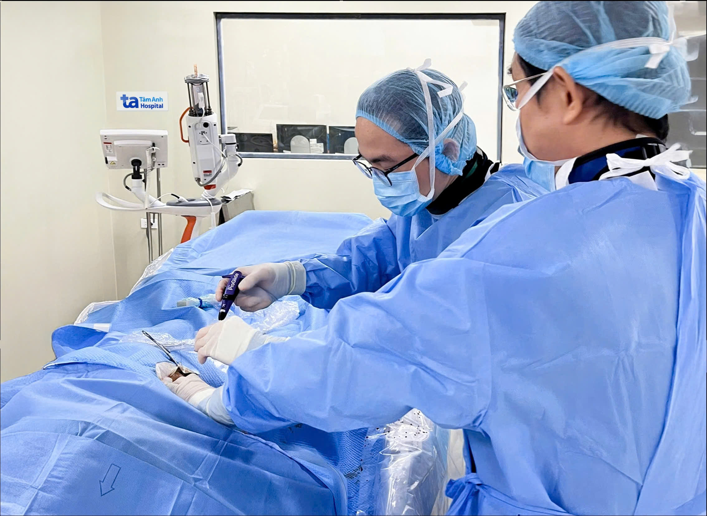
After consultation, the medical team decided to address the appendiceal mucocele first. The biopsy confirmed it was benign. Two months later, after a full recovery, Tin was ready for the aortic aneurysm procedure.
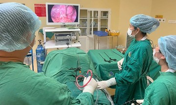
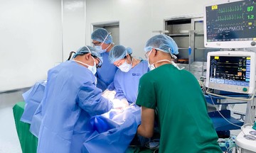
Dr. Dung opted for a less invasive stent graft placement, inserting a covered stent to redirect blood flow away from the weakened section of the aorta. This approach minimized procedure time, offered high treatment efficacy, reduced complication risks, and promoted faster recovery. The team inserted a catheter through Tin's femoral artery, guiding it to the affected area in the thoracic aorta, and then deployed the stent graft. Tin's post-operative recovery was smooth.
 |
Doctors place a stent graft to treat the patient's aortic aneurysm. Photo: Ha Vu |
Doctors place a stent graft to treat the patient's aortic aneurysm. Photo: Ha Vu
Doctor Tran Quoc Hoai of Tam Anh General Hospital's Department of Cardiothoracic and Vascular Surgery emphasized the danger of aortic aneurysms, which can be life-threatening if left untreated. The aorta, the body's largest artery, carries oxygenated blood from the heart. A weakened aortic wall can form an aneurysm, a bulge that disrupts blood flow and can lead to serious complications.
Abdominal aortic aneurysms are the most common type (75%), followed by thoracic aortic aneurysms, which occur in the chest area. Men over 65, long-term smokers, those with a family history of aortic aneurysms, and individuals with hypertension are at higher risk.
A ruptured aneurysm is a life-threatening emergency. The resulting internal bleeding can cause severe hemorrhagic shock, depriving vital organs like the heart, brain, and kidneys of blood. Without immediate intervention, the risk of death is very high.
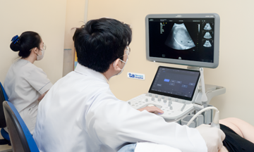
According to Dr. Hoai, most aortic aneurysms are asymptomatic in the early stages, making detection difficult. Untreated, the aneurysm continues to grow, increasing the risk of rupture. The condition often progresses silently, discovered incidentally through imaging tests like abdominal ultrasounds for initial screening and CT angiography (CTA) for precise sizing, location, and treatment planning.
Individuals experiencing symptoms suggestive of an aortic aneurysm, such as persistent dull abdominal or back pain, chest pain radiating to the back, chronic cough, hoarseness, or shortness of breath, should seek immediate medical attention. Signs of a ruptured aneurysm include pale skin, cold extremities, sweating, dizziness, fainting, rapid heartbeat, nausea and vomiting, shortness of breath, and sudden, escalating pain in the abdomen, lower back, or legs. These symptoms require immediate emergency care.
Thu Ha
* The patient's name has been changed.
| Readers can submit questions about cardiovascular diseases here for doctors to answer. |