On 5/8, Doctor Nghiem Thi Hong Hanh from Hitec Eye Hospital, highlighted the importance of retinal vascular examinations in early detection of stroke and sudden cardiac death risks. This non-invasive method allows doctors to "see" the health of the entire vascular system through the "retinal window". This connection is based on the principle that the retinal vasculature shares a similar structure and is influenced by the same risk factors as the vasculature in the brain and heart.
Changes in retinal blood vessels, such as narrowing, dilation, bleeding, or small blockages, accurately reflect the condition of the small blood vessels in the brain and heart. Conditions like hypertension, diabetes, high cholesterol, and atherosclerosis, leading risk factors for stroke and heart attacks, often cause early damage to the retinal microvasculature.
Hypertensive retinopathy causes hardening of the vessel walls (copper wire sign), retinal hemorrhages, cotton wool spots, and optic disc swelling. Diabetic retinopathy causes microaneurysms, dot and blot hemorrhages. Blood clots or atherosclerotic plaques can become lodged in retinal blood vessels, causing "eye stroke" (retinal artery occlusion).
"This is one of the warning signs of an impending stroke or heart attack," Dr. Hanh explained. Therefore, retinal vascular examination not only helps diagnose eye diseases but also assesses the health of the entire vascular system, especially the brain and heart. Early detection of damage from hypertension, diabetes, and atherosclerosis provides warning signs of a high risk of stroke and cardiovascular disease in the near future.
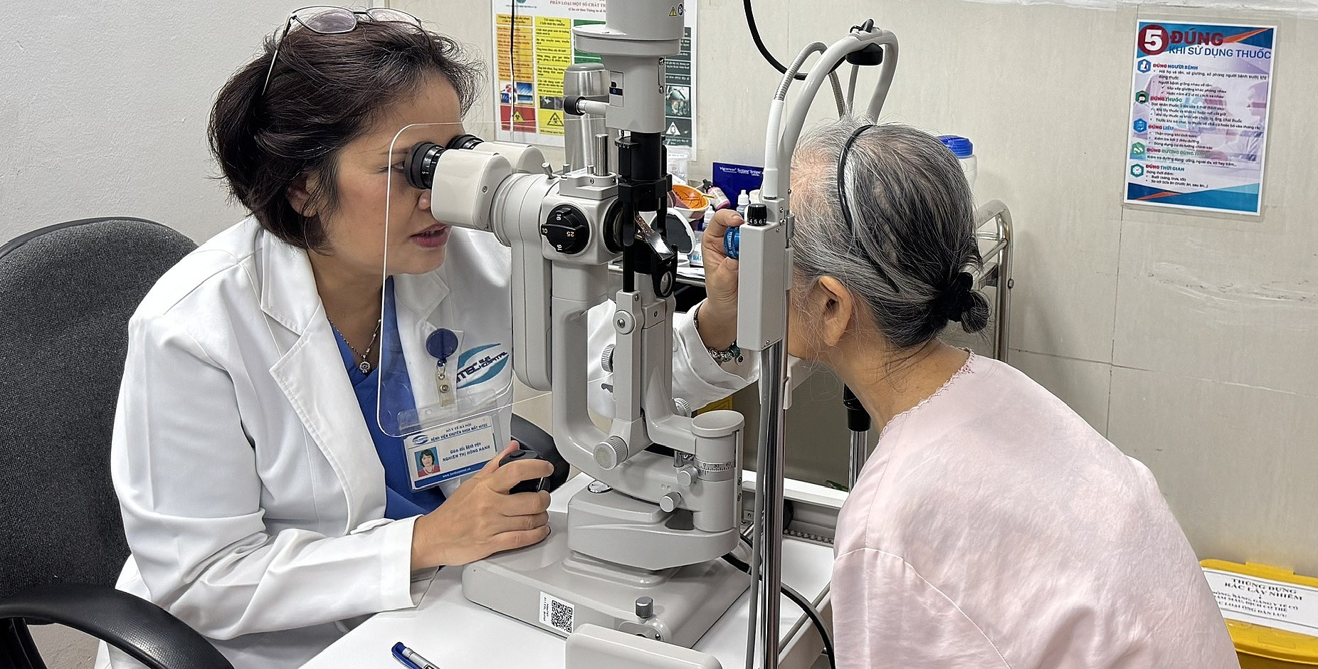 |
A doctor examines a patient's eyes. Photo: Hospital provided |
A doctor examines a patient's eyes. Photo: Hospital provided
Dr. Hanh said that the hospital has integrated AI technology to screen for and detect early retinal damage, as well as some chronic diseases that cause microvascular damage.
Specifically, the retinal imaging software uses a camera combined with an AI algorithm. This algorithm analyzes images of the microvasculature, helping to detect the risk of diseases such as glaucoma, macular degeneration, neovascularization, and diabetic retinopathy with up to 95% accuracy.
"In the field of cardiology, the system also provides early warning indicators of disease risk. These indicators are analyzed from the state of the retinal microvasculature recorded in fundus photographs," Dr. Hanh added.
Sudden cardiac death and stroke are two dangerous medical conditions that can cause death or permanent disability. Recently, the frequency of sudden cardiac death and stroke has been increasing alarmingly, not only in the elderly but also in seemingly healthy young people.
Sudden cardiac death is an unexpected death that occurs within a short period, usually within an hour of symptom onset. Stroke is a condition in which the brain is severely damaged due to lack of oxygen. There are two types of stroke: ischemic (caused by a blockage) and hemorrhagic (caused by bleeding).
Experts recommend a 5-10 minute eye exam to examine the retinal vasculature for assessing brain and heart health and detecting potential abnormalities even before clinical manifestations appear. Patients are advised to consult with a cardiologist to rule out risks early and proactively develop a personal health care plan.
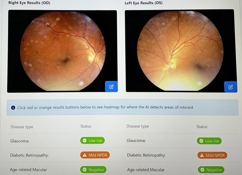 |
Image analyzed by AI warning of the risk of retinal damage due to diabetes. Photo: Hospital provided |
Image analyzed by AI warning of the risk of retinal damage due to diabetes. Photo: Hospital provided
Le Nga