Ms. Them underwent a specialized cardiac CT scan at Tam Anh General Hospital, Ho Chi Minh City, revealing partial anomalous pulmonary venous return (PAPVR) with a sinus venosus atrial septal defect. Doctor Vu Nang Phuc, Head of the Congenital Heart Department at the Heart Center, stated this is a rare form of congenital heart defect in adults.
Normally, oxygen-rich blood returns from the lungs to the heart via four pulmonary veins, emptying directly into the left atrium. However, in Ms. Them's case, the right upper pulmonary vein "went astray," not emptying into the left atrium as usual but connecting abnormally to the superior vena cava. This phenomenon is known as partial anomalous pulmonary venous return.
PAPVR in the right upper pulmonary vein region often co-occurs with a sinus venosus atrial septal defect. Ms. Them also had a small defect in the upper region between the right and left atria, which is of this type. These two congenital abnormalities appearing together meant oxygen-rich blood from the lungs could not reach the left heart but instead flowed back to the right heart. This condition overworks and enlarges the right heart chambers, increasing blood flow to the lungs and leading to progressive pulmonary hypertension. Without timely detection and treatment, patients can develop right heart failure, leg swelling, cyanosis of the lips, or dangerous arrhythmias.
Ms. Them previously had a dual-chamber pacemaker implanted due to sick sinus syndrome, a type of heart rhythm disorder. According to Doctor Phuc, the pacemaker leads were positioned close to the anomalous connection, obscuring images on echocardiograms and making misdiagnosis or overlooking the underlying defect easy.
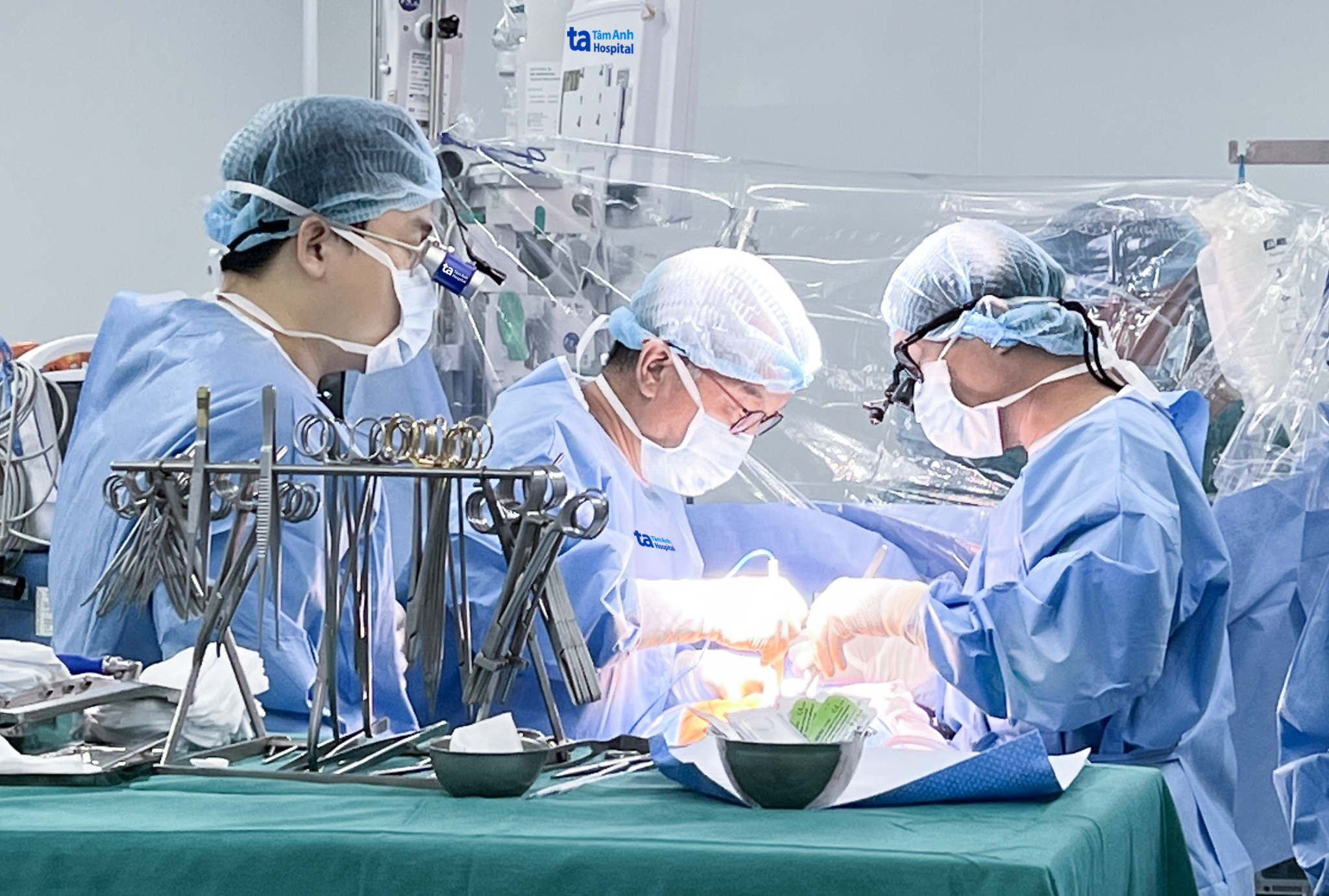 |
Surgeons repair the heart's blood vessels for the patient. *Photo: Minh Huyen* |
The surgical team created a tunnel in the heart to redirect blood flow to the correct left atrial chamber. Master of Science, Doctor Nguyen Minh Tri Vien, a cardiac surgery consultant at Tam Anh General Hospital, Ho Chi Minh City, stated this surgery carried many risks because the pacemaker leads were located directly at the anomalous connection. Without careful planning, dissection could sever or displace the leads, causing the patient to lose rhythm and require another pacemaker implantation.
For over 5 hours, surgeons temporarily paused the pacemaker, opened the right heart and superior vena cava to create a tunnel, redirecting blood from the pulmonary veins to the left atrium. Simultaneously, the team preserved natural flow pathways to prevent future narrowing.
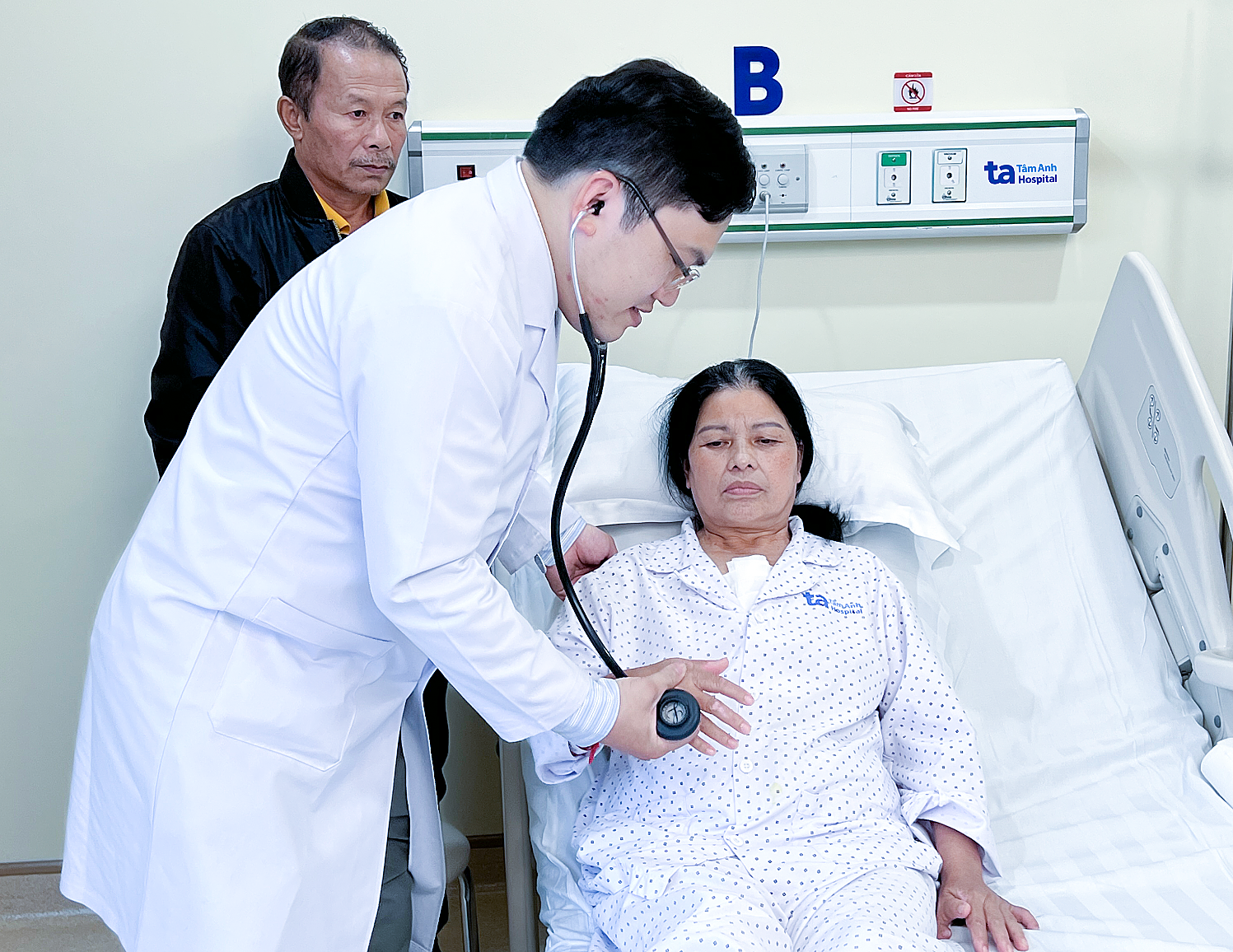 |
Doctor Phuc examines the patient before discharge. *Photo: Minh Huyen* |
Post-surgery, Ms. Them's pacemaker was safely checked and reactivated. She no longer felt fatigued or short of breath and was discharged after 7 days. Her right heart chambers had contracted, pulmonary artery pressure returned to near normal, and the electrodes were functioning stably.
According to Doctor Phuc, without a detailed cardiac CT scan, doctors could easily miss the defect or misdirect treatment. Partial anomalous pulmonary venous return with a sinus venosus atrial septal defect might not show symptoms for many years. As patients age, they can easily mistake fatigue and shortness of breath for lung disease or hypertension, leading to delayed diagnosis. In patients with pacemakers, images inside the heart can be obscured or distorted. Therefore, patients should seek early medical attention if they experience persistent unusual symptoms.
Thu Ha
| Readers can submit questions about cardiovascular diseases here for doctors to answer |