Doctors at Benh vien Da khoa Tam Anh Ha Noi discovered a tumor in the patient's lower abdomen that extended above her navel. An ultrasound revealed a right ovarian tumor measuring 185x96x132 mm, containing multiple septations and a solid component measuring 43x41 mm with minimal vascularization on Doppler. The tumor was classified as ORADS 4, indicating a high risk of malignancy.
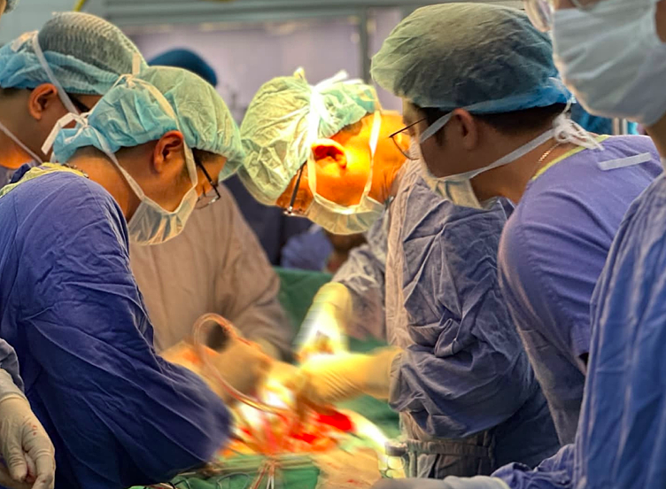
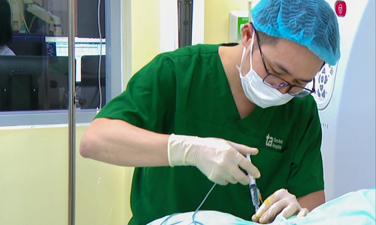
Doctor Nong Tuyet Phuong, from the Center for Obstetrics and Gynecology, prescribed laparoscopic surgery to remove the tumor and for histopathological evaluation. The medical team inserted a trocar (a surgical instrument for laparoscopy) through the navel directly into the tumor, aspirating approximately one liter of clear yellow fluid. After insufflating CO₂ gas and inserting a camera into the abdominal cavity, doctors noted that the inner and outer surfaces of the tumor were relatively smooth, and inside, there was a papilla approximately 3 cm in size.
The doctor dissected and completely removed the right ovarian tumor, which weighed about 2 kg, while maximally preserving healthy reproductive structures. The specimen underwent frozen section biopsy during surgery. Results confirmed the tumor contained various tissue types originating from all three germ layers (ectoderm, mesoderm, endoderm), such as skin, hair, nerve tissue, fat, muscle, bone, cartilage, and glandular epithelium.
Based on histopathological criteria, Doctor Phuong diagnosed an immature teratoma, grade one—a type of malignant ovarian germ cell tumor.
Post-surgery, Phuong received antibiotics, anti-inflammatory medication, and nutritional support. Because the tumor was detected at an early stage, localized to the ovary, and completely removed surgically, the patient did not require chemotherapy.
According to Doctor Phuong, cases of immature teratoma, grade one, that are detected early and treated thoroughly, generally have a favorable prognosis. Patients need regular follow-up examinations to detect any risk of recurrence early.
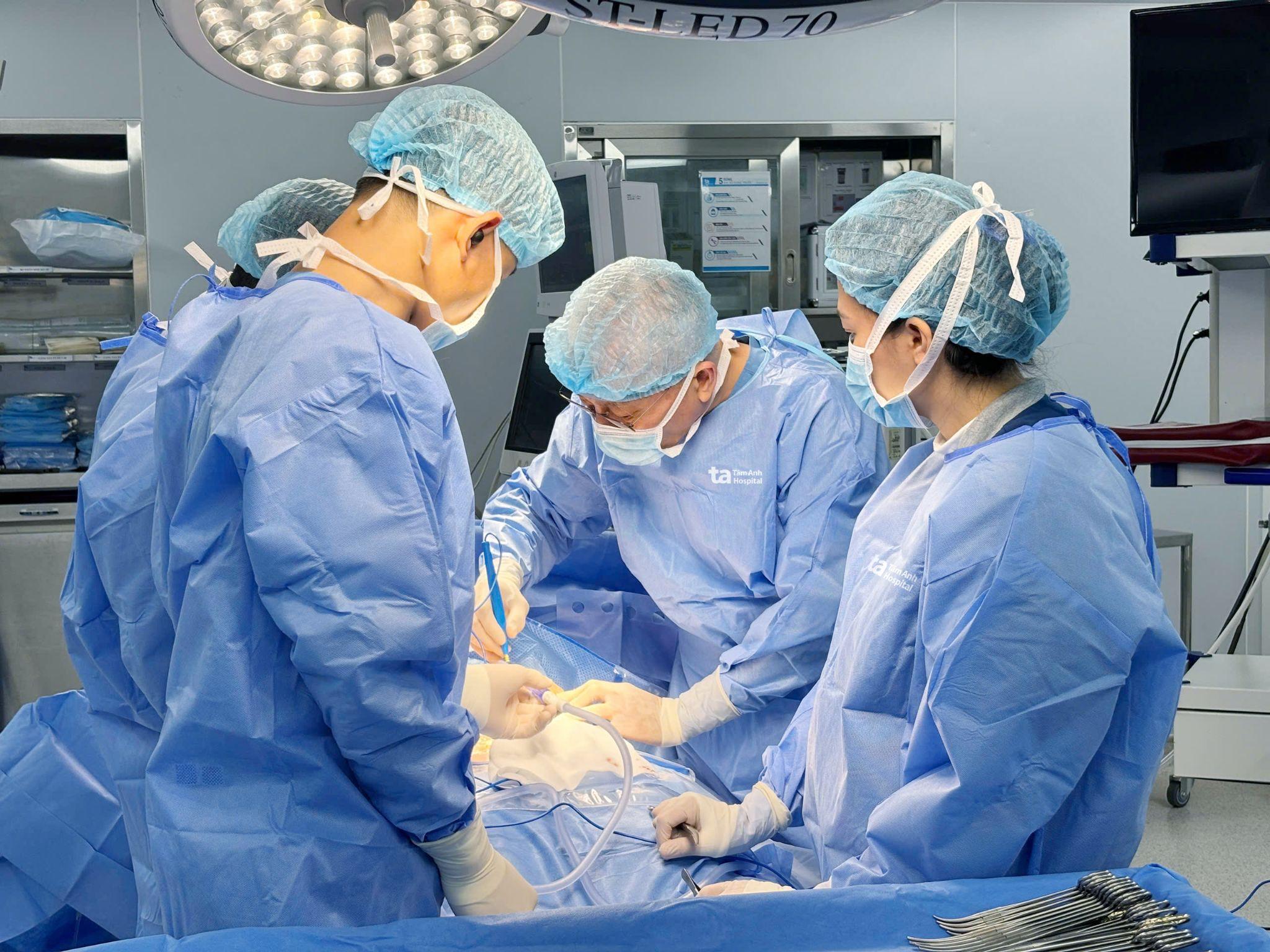 |
The medical team from the Center for Obstetrics and Gynecology performing surgery on a patient. Illustration: Benh vien Da khoa Tam Anh
Ovarian teratomas originate from ovarian germ cells. These cells can differentiate into various tissue types in the body. When development and differentiation occur abnormally, these cells can form a tumor containing hair, skin, teeth, bone, cartilage, or nerve tissue. Because they arise from abnormal germ cell development, the condition can appear at any age. Depending on the maturity level of the internal tissue components, tumors are classified as mature or immature teratomas.
Mature teratomas constitute the majority and are typically benign. Doctor Phuong cited studies showing that immature teratomas account for less than 1% of all ovarian cancers.
The disease often progresses silently with non-specific symptoms such as bloating, gradual abdominal enlargement, lower abdominal pain, a palpable mass in the abdomen, or menstrual irregularities. Consequently, many cases are only discovered when the tumor has grown to a large size.
Doctor Phuong advises young women, including those who are unmarried or not sexually active, not to be complacent if they experience symptoms like abnormal abdominal enlargement, prolonged abdominal pain, or a heavy, dull sensation in the lower abdomen. Seeking medical examination helps detect ovarian tumors early before complications such as torsion, cyst rupture, or progression to malignancy occur.
Trinh Mai
*The patient's name has been changed.
| Readers can submit questions about pregnancy and childbirth here for doctors to answer |