Quynh, 31 years old and pregnant with her second child, discovered a true umbilical cord knot during a Doppler ultrasound at week 32 at Tam Anh General Hospital Hanoi. Dr. Duong Ngoc Hung, from the OB/GYN Center, stated that because the fetus continued to receive oxygen and nutrients and developed appropriately for its gestational age, immediate intervention was not necessary.
Doctors monitored Quynh using Doppler ultrasound to assess blood flow through the umbilical cord and the fetal cerebral circulation, combined with computerized cardiotocography (cCTG). This method acts as a "radar" to precisely analyze short-term variation (STV), a critical indicator of the fetal central nervous system's health.
cCTG identifies and classifies early decelerations in the fetal heart rate. The monitoring results provide additional data for doctors to evaluate the fetal condition and determine the appropriate time for intervention.
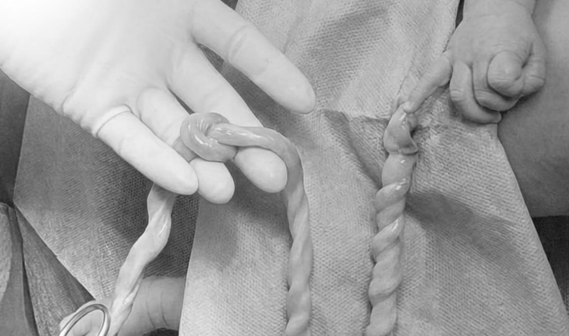 |
The baby's umbilical cord with one loop. *Tam Anh General Hospital*. |
Dr. Hung explained that unlike traditional cardiotocography (CTG), which relies on visual interpretation, cCTG applies computer algorithms (most commonly the Dawes-Redman system) to analyze fetal heart rate indicators. The system automatically analyzes fetal heart rate data and uterine contractions every 1/16 minute, equivalent to 3,75 seconds, standardizing data and reducing errors when multiple observers are involved.
After five weeks of monitoring, doctors scheduled a proactive C-section at week 37 to mitigate the risk of the knot tightening during the final stage of pregnancy or during labor. The baby boy was born weighing 2,9 kg, in stable health, and was discharged after four days.
 |
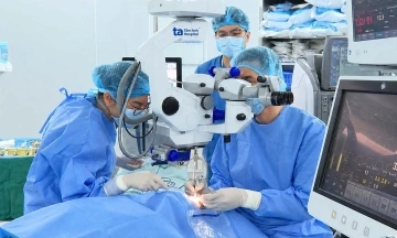
Doctor Hung performing a C-section for the pregnant woman. *Tam Anh General Hospital*. |
Dr. Hung noted that true umbilical cord knots occur in approximately 0,3-2% of pregnancies. The knot in the umbilical cord can tighten when the fetus moves or during labor, interrupting blood flow to the fetus. This can lead to fetal distress, birth asphyxia, or stillbirth. Factors that may increase this risk include: a long umbilical cord, polyhydramnios, active fetal movement, post-term pregnancy, gestational diabetes, chronic hypertension, having given birth multiple times, or a history of miscarriage.
True umbilical cord knots often present no specific symptoms. Many cases are only detected through ultrasound, or even during a C-section or after birth. Color Doppler ultrasound helps identify umbilical cord anomalies and assess blood flow through the vessels.
Upon detection or suspicion of a true umbilical cord knot, doctors assess the gestational age, fetal development, amniotic fluid volume, cord blood flow, fetal heart rate, and fetal movement. If the fetus remains stable with no signs of circulatory impact, monitoring can continue with cCTG.
Dr. Hung advises pregnant women to attend scheduled prenatal check-ups, paying particular attention to fetal movements in the later weeks. If fetal movements decrease significantly or become notably weak, accompanied by abdominal pain, bleeding, or vaginal fluid leakage, prompt medical attention is necessary.
Thuy Hanh
| Readers can submit questions about obstetrics and gynecology here for doctors to answer. |