Ms. Nam, 86, experienced increasingly frequent chest pains in recent months. With a history of hypertension and type 2 diabetes, she had two stents placed in her right coronary artery in 2019. Master of Science, Doctor Duong Cong Linh, from the Cardiology Department at Tam Anh General Hospital Hanoi, diagnosed her with unstable angina due to multi-branch coronary artery disease. A computed tomography scan revealed that while her two existing stents remained clear, her left anterior descending artery (LAD) was severely narrowed by 80-90% in multiple segments.
The LAD is the main branch supplying blood to most of the heart's anterior wall and interventricular septum. When this vessel significantly narrows, reduced blood flow to the heart muscle causes oxygen deprivation, leading to angina. Without timely revascularization, atherosclerotic plaques at the narrowed site can progress or rupture, forming blood clots that completely block the vessel and lead to myocardial infarction.
A multi-slice computed tomography scan showed a tightly narrowed arterial segment with thick, hard calcified plaque encasing the vessel wall. While a normal blood vessel resembles a soft rubber tube, calcification makes it rigid like a plastic tube. This type of damage makes it difficult for conventional balloon catheters to expand optimally, and stents struggle to fully appose the vessel wall, increasing the risk of restenosis or complications.
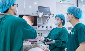
According to Doctor Linh, complex cases previously often required coronary artery bypass graft (CABG) surgery. Although effective, this method carries significant risks, especially for elderly patients or those with underlying conditions like hypertension, diabetes, or kidney failure. Currently, the optimal solution is intravascular lithotripsy (IVL), a technology that treats calcified plaques within the coronary arteries using shockwaves.
IVL effectively "softens the concrete base" before stent placement, allowing the stent to expand maximally and appose well. A specialized IVL balloon is advanced to the narrowed site, emitting high-energy mechanical pulses that directly impact the calcified plaque. These pulses fracture surface calcium and layers deep beneath the intima, making the vessel wall softer and more expansive, with minimal impact on surrounding soft tissue.
Following the IVL procedure, the medical team placed two drug-eluting stents in the narrowed segments of the LAD, restoring blood flow to the myocardium. The entire process was monitored using intravascular optical coherence tomography (OCT), which allowed for precise assessment of calcium fracture and stent apposition.
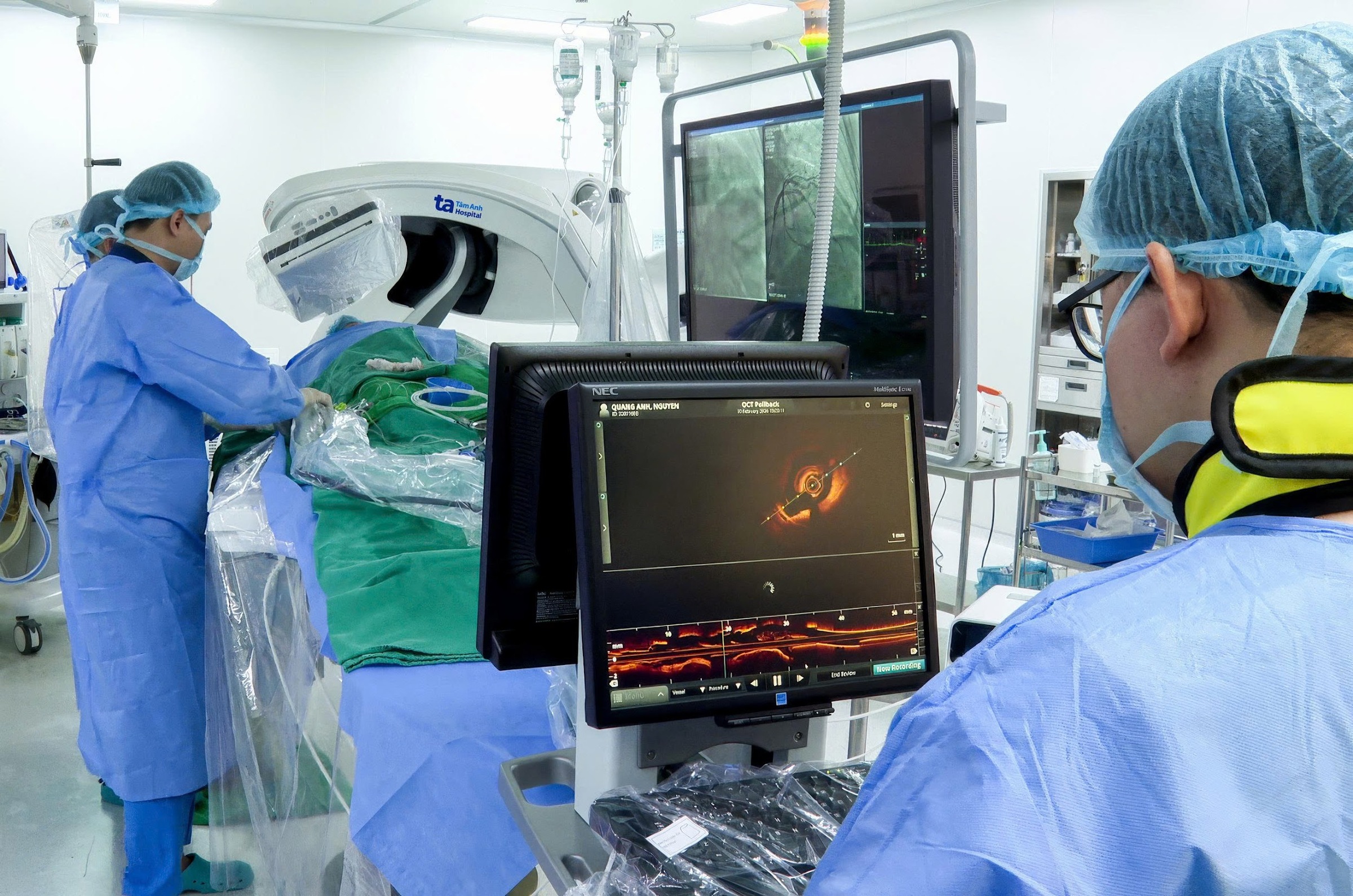 |
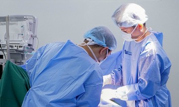
The intervention team breaks down coronary artery calcification using IVL technology and places stents to restore blood flow. *Photo: Tam Anh General Hospital*.
After the intervention, Ms. Nam no longer experienced chest pain or shortness of breath, and her health stabilized. Following discharge, she was scheduled for regular follow-up appointments, where doctors continue to monitor her and provide medical treatment to control her blood pressure and blood sugar.
Doctor Linh noted that IVL technology is widely applied in many major cardiac centers in the United States and Europe. This method improves success rates when placing stents in severe calcified lesions, while also ensuring safety and minimal invasiveness. In Vietnam, IVL has only recently been implemented.
Ly Nguyen
*Patient's name has been changed.
| Readers can submit questions about cardiology here for doctors to answer. |