About three years ago, Ngan noticed a small, painless lump, about the size of a fingertip, on his forehead. As it grew, he consulted a doctor and was diagnosed with a tumor. However, feeling otherwise healthy, he opted not to pursue treatment. Over the past year, the tumor grew significantly, causing pressure when he bent over or lay on his side. Still, he attributed the symptoms to minor issues.
Dr. Chu Tan Si, Head of Neurosurgery at Tam Anh General Hospital in Ho Chi Minh City, explained that Ngan initially came to the hospital for back pain and a herniated disc. During the examination, doctors discovered the unusual protrusion on his left forehead, extending towards his left temple and ear.
A 3 Tesla MRI scan revealed a large brain tumor, approximately 11cm in diameter, attached to the dura mater and extending into the soft tissue beneath the skin of his forehead. The tumor had eroded the frontal and parietal bones of his skull, compressed the left frontal lobe, and displaced the midline of his brain by almost 1cm. It also caused a slight herniation of the left uncus through the tentorium cerebelli, a warning sign of potential brain herniation.
Dr. Si explained that brain tumors exceeding 5-6cm typically cause brain swelling, compression of functional areas, and brainstem herniation. Ngan hadn't experienced typical symptoms like headaches, nausea, papilledema, or weakness because the tumor was located in the frontal lobe—an area of the brain less associated with sensory or motor functions. “The tumor's growth outside the skull, eroding the frontal and parietal bones, inadvertently created an escape route, temporarily relieving intracranial pressure," Dr. Si said. He added that despite this, surgery was crucial due to the tumor's unusual size, its impact on the skull base, and the threat to the brainstem, which posed a constant risk of brain swelling and herniation.
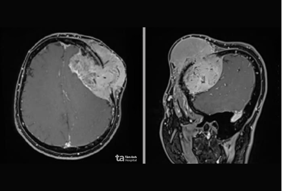 |
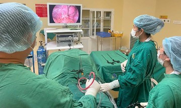
An MRI scan shows the large tumor protruding from Ngan's left forehead. Photo: Tam Anh General Hospital |
An MRI scan shows the large tumor protruding from Ngan's left forehead. Photo: Tam Anh General Hospital
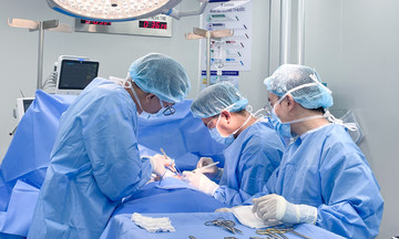
Doctors decided to surgically remove the tumor using neuronavigation and an AI-integrated surgical microscope. The incision followed a curved path through Ngan’s hairline to minimize scarring. Due to the tumor’s vascularity and risk of bleeding, doctors meticulously cauterized and cut each feeding vessel from the dura mater and arachnoid mater. They then employed the "debulking" technique, using a Cusa ultrasonic aspirator to reduce the tumor's volume before dissecting the edges, minimizing potential damage to surrounding brain tissue. Portions of the tumor adhered to the cerebral cortex were carefully removed using microsurgical instruments.
After nearly three hours, the entire tumor was successfully removed in sections, with no damage to the cerebral cortex or venous sinuses, and Ngan's neurological functions preserved. Doctors reconstructed his skull using a specialized titanium implant, a common procedure in neurosurgery for large skull defects to restore the skull's shape and protect the brain.
 |
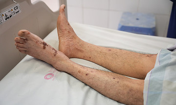
Dr. Si examines Ngan's incision after the surgery. Photo: Tam Anh General Hospital |
Dr. Si examines Ngan's incision after the surgery. Photo: Tam Anh General Hospital
Ngan recovered quickly, experiencing only mild numbness at the incision site. He was walking, eating, and communicating normally within a day and was discharged after 7 days. He plans to address his herniated disc after two months of recovery. A biopsy confirmed the tumor as a benign meningioma, a type that grows slowly and often goes unnoticed until it reaches a significant size.
Doctors advise against ignoring unusual lumps or signs in the head and neck, even if painless. Any such symptoms warrant immediate consultation with a neurologist for diagnosis and timely intervention.
Lan Anh
*The patient's name has been changed.
| Readers can submit questions about neurological conditions here for doctors to answer. |