Khuyen's computed tomography (CT) scan at Tam Anh General Hospital in Ho Chi Minh City revealed a large mass extending into the eye socket, compressing the eyeball and surrounding structures. Professor Tran Phan Chung Thuy, consultant at the ENT Center, diagnosed her with recurrent rhinosinusitis, frontal sinus mucocele, and a deviated septum. Surgery was recommended to remove the inflamed tissue and mucocele, and to improve sinus ventilation. However, Khuyen's previous sinus surgery and mucocele removal meant her sinus anatomy was altered, increasing the risk of surgical complications.
Professor Thuy explained that revision sinus surgery is often more complex than the initial procedure. The sinus structure is changed, and scar tissue obscures natural anatomical landmarks. Differentiating between diseased and healthy tissue becomes challenging, making dissection more precarious. Incomplete removal of inflamed tissue can lead to recurrence. The proximity of the sinuses to critical structures like the eye socket, skull base, optic nerve, and internal carotid artery demands precise surgical maneuvers within a confined space. Even minor surgical errors can result in complications such as reduced vision, permanent vision loss, cerebrospinal fluid leaks, facial paralysis, or massive bleeding.
The surgical team utilized an AI-powered image-guided surgery (IGS) system combined with endoscopy, 3D imaging, and 4K resolution to pinpoint the mass's location and remove the diseased tissue while minimizing complications and shortening the surgery time. Before the procedure, Khuyen's CT scan was uploaded into the IGS system to synchronize data, reconstruct the sinus space, and display it on a screen.
After anesthesia, a sensor was attached to a fixed point on Khuyen's forehead. The AI technology performed a 3D scan, recognizing her facial structure and comparing it with the preoperative CT data. Throughout the surgery, the AI displayed the surgical instruments' trajectory on the reconstructed 3D sinus "map" on the screen, providing warnings if they approached hazardous areas.
The system synchronized Khuyen's real-time surgical anatomy with the CT/MRI images on the screen, allowing the surgeon to view both simultaneously with the endoscopic image. In conventional endoscopic surgery, surgeons might have to pause to consult CT/MRI images on a separate display to confirm their position.
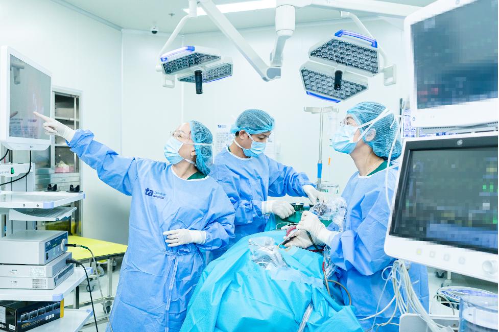 |
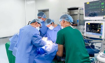
Professor Thuy (left) and the surgical team operating on the patient. Photo: Tam Anh General Hospital |
Professor Thuy (left) and the surgical team operating on the patient. Photo: Tam Anh General Hospital
Professor Thuy widened the frontal sinus drainage pathway, creating access to the mucocele. Guided by the IGS system, the entire mass was successfully removed without damaging healthy tissue. Simultaneously, the inflamed tissue was addressed, and pus drainage from the frontal sinus was facilitated.
The successful surgery concluded almost an hour earlier than anticipated. Khuyen recovered well postoperatively and was discharged after a week.
Uyen Trinh
| Readers can submit questions about ENT diseases here for doctor's answers. |