Ms. Tham, 40 years old, was 29 weeks pregnant when the fetus showed signs of being underweight. At Tam Anh General Hospital, Ho Chi Minh City, Master of Science, Doctor La Hong Chau, Head of the Obstetrics and Gynecology Imaging Unit at the Center for Diagnostic Imaging and Interventional Radiology, performed a 4D morphological ultrasound. The ultrasound revealed a small fetus and a parasitic twin mass on the fetus's buttocks. This parasitic mass had no heart, head, or internal organs, only skeletal structures and soft tissue, receiving blood supply from the main fetus.
Based on Ms. Tham's history of a natural twin pregnancy and the characteristics of the abnormal mass seen on ultrasound, Dr. Chau diagnosed it as a parasitic twin. Dr. Chau explained that the orderly arrangement of long bones and underdeveloped limb buds are key signs distinguishing it from a sacrococcygeal teratoma. Parasitic twins occur when monozygotic twins develop abnormally from an early stage. One twin ceases development but continues to receive blood from the other, existing as a body part attached to the main fetus.
"World medical literature records dozens of cases of parasitic twins in the buttock-sacrococcygeal region," Dr. Chau said.
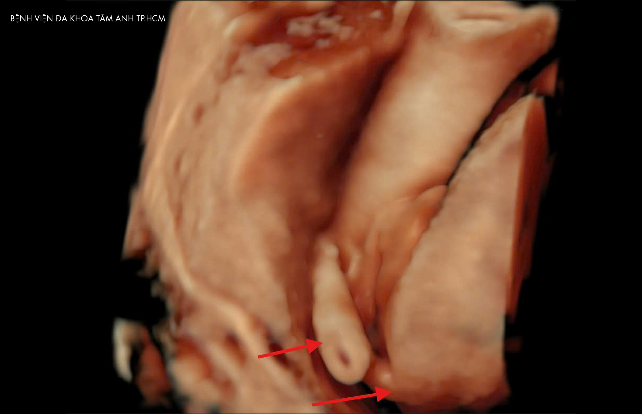 |
Fetal MRI results showing the parasitic twin mass in the buttock area. Photo: Tam Anh General Hospital |
Upon discovering the parasitic twin, Ms. Tham's family chose to continue monitoring the pregnancy. Doctors prescribed ultrasound check-ups every two weeks. Doctor, Specialist Level 1 Tran Lam Khoa, from the Obstetrics and Gynecology Center at Tam Anh General Hospital, Ho Chi Minh City, noted that while the main fetus was small for its gestational age, the parasitic twin mass did not increase in size and had not yet affected the fetus's development, so no in-utero intervention was needed. Dr. Lam Khoa advised Ms. Tham to increase her nutritional intake during pregnancy, allowing the main fetus to catch up to a normal growth rate.
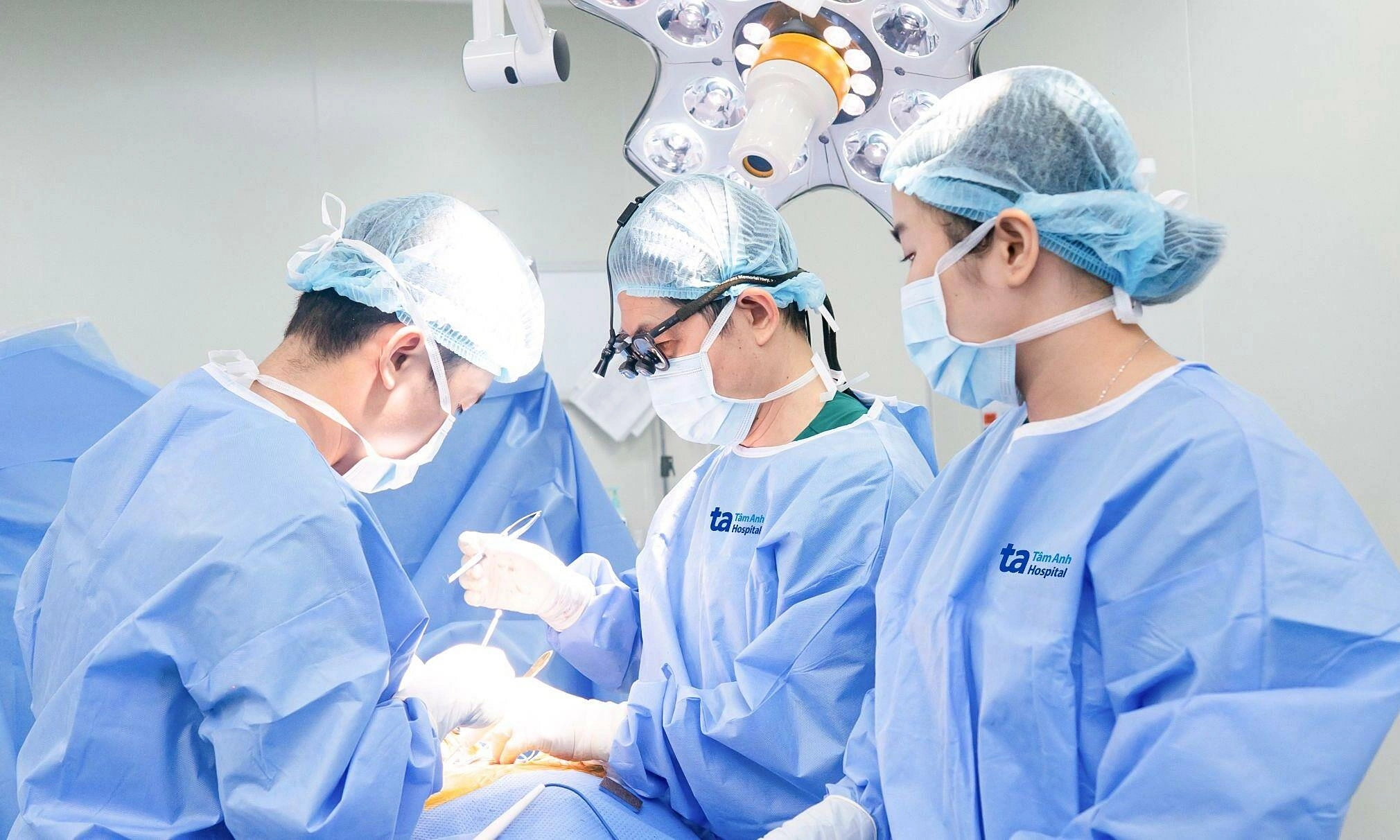
At 38 weeks, Ms. Tham went into labor. Doctors opted for a C-section due to a prior surgical scar. A healthy baby girl was born weighing 3.2 kg, with a coin-sized round soft tissue mass on her left buttock and a 4 cm long tissue mass in the coccyx region.
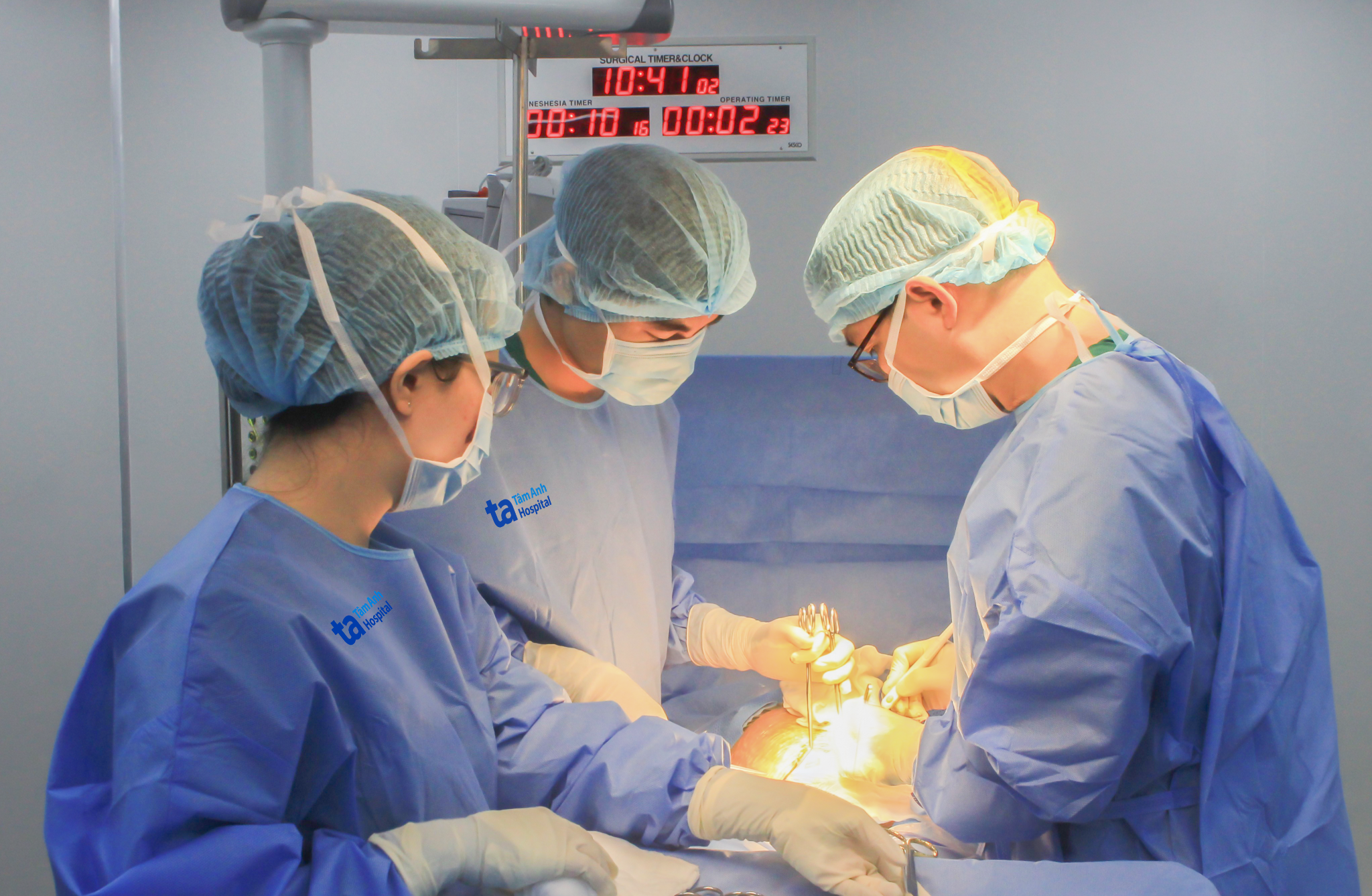 |
Doctor, Specialist Level 2 Le Thanh Hung (right) and his team performing a C-section for Ms. Tham. Photo: Tam Anh General Hospital |
One day after birth, the baby underwent an MRI to precisely determine the size of the parasitic mass, its internal structure, blood supply, and its relationship with the baby's spine, spinal cord, blood vessels, and adjacent organs. Doctor, Specialist Level 1 Le Thi Ngoc Dung, from the Neonatal Center, stated that the results were consistent with the prenatal diagnosis. The parasitic twin mass had not caused compression, circulatory disorders, infection, or affected organ function, so surgery was not immediately necessary.
The baby girl was discharged with her mother and will have regular follow-up appointments to determine the optimal time for intervention.
According to Dr. Lam Khoa, there is currently no standard international classification system for parasitic twins in medical literature. Parasitic twins are classified based on their attachment site to the main fetus. There are five to six main types recorded, including parasitic twins of the buttock-sacrococcygeal region, abdominal wall, chest, pelvis, head, and spine.
Most parasitic twins are detected during pregnancy through ultrasound and MRI. Early detection of birth defects in utero helps doctors plan monitoring, choose the timing of birth, and determine appropriate post-birth interventions for the child.
Tue Diem
*Patient's name has been changed
| Readers can submit questions about pregnancy and childbirth here for doctors to answer |