Professor, Doctor Vo Thanh Nhan, Director of the Interventional Cardiology Center at Tam Anh General Hospital, Ho Chi Minh City, stated that in-stent restenosis is a complication following coronary stent placement. It occurs when the vessel lumen narrows again after being expanded by a stent. Mrs. Tam experienced 45-50% in-stent narrowing in her left anterior descending artery and 40-50% in the remaining branch. This condition led to myocardial ischemia, causing her persistent chest pain with increasing frequency.
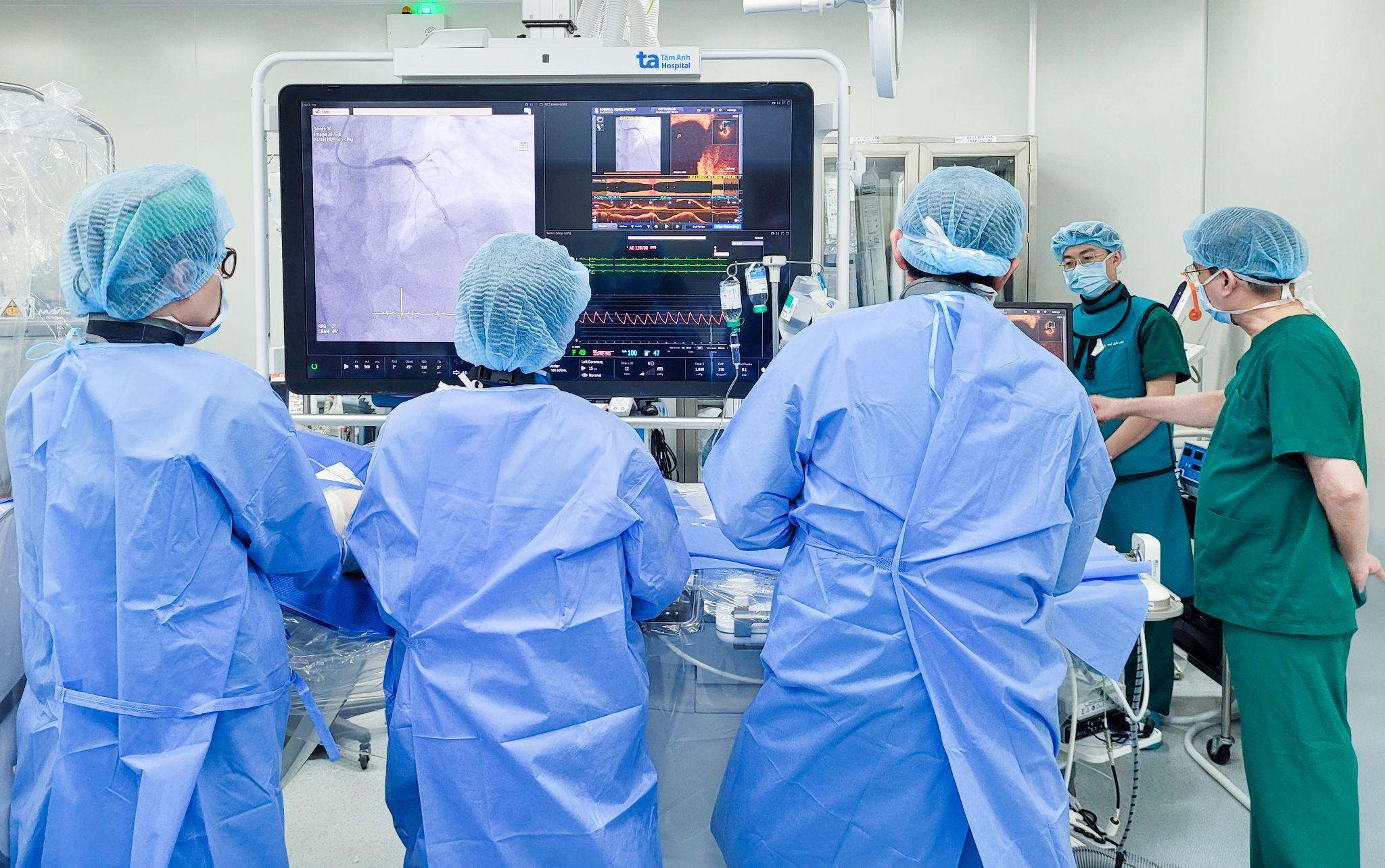
A coronary CT scan indicated that the degree of narrowing was not severe. However, according to Professor Nhan, this is only a two-dimensional X-ray image of the vessel lumen, which does not reflect the vessel wall structure or accurately determine the extent of restenosis. Therefore, the patient underwent optical coherence tomography (OCT), which uses infrared light to create detailed, high-resolution images (10-20 microns) of the coronary artery lumen. This allowed doctors to clearly observe the thickness, composition, and structure of atherosclerotic plaques, as well as the degree of in-stent restenosis, aiding in the selection of an appropriate stent.
 |
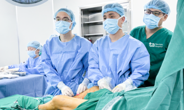
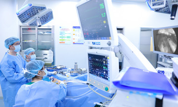
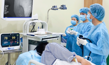
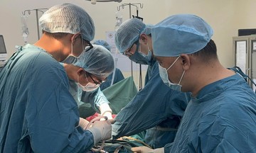
Doctors place a stent to re-open the coronary artery for a patient. Photo: Ha Vu |
Mrs. Tam's OCT results revealed that the damage was far more severe than indicated by the CT images. Her left anterior descending artery showed 70-90% restenosis both within and after the stent, the circumflex artery had 95-99% in-stent restenosis, and the right coronary artery had 50% restenosis.
The patient's cardiac vascular system showed severe narrowing in multiple locations, accompanied by atherosclerosis and calcification, posing a high risk of incomplete revascularization after intervention. The left anterior descending artery, a main blood supply to the heart, is located close to the heart pedicle, making stent insertion inherently risky. Incorrect guidewire manipulation could damage the heart, leading to danger. The medical team decided to perform angioplasty and stenting with the support of OCT technology.
After 30 minutes, doctors successfully placed two stents, measuring 2.5x38 mm and 2.75x38 mm, to fully cover the damaged areas within and beyond the two old stents. Doctors also checked the stent edges to rule out complications like dissection, which could cause acute in-stent thrombosis. Once coronary blood flow was re-established, Mrs. Tam's chest pain significantly decreased, and she was discharged two days later in stable condition.
According to Doctor Duong, in-stent restenosis is a major challenge after intervention, affecting approximately 5-15% of all cases. Restenosis within 12 months of stent placement is often due to incorrect stent sizing or suboptimal stenting technique, where the stent does not fully expand or does not tightly adhere to the vessel wall. If a patient experiences narrowing again after one year, the primary cause is often atherosclerosis. To reduce the risk of restenosis, patients must strictly adhere to medication regimens, control risk factors through a healthy lifestyle such as quitting smoking, limiting alcohol, exercising regularly, maintaining a good diet, and managing underlying conditions.
Thu Ha
*Patient's name has been changed
| Readers can send their cardiology questions here for a doctor's response |