Response:
Cochlear implant surgery is complex due to the small, narrow structures within the ear, which contain numerous blood vessels and nerves. An AI-powered surgical microscope magnifies images and provides focused light, allowing surgeons to clearly view the surgical field and detailed anatomical structures. This precision guides each step, such as opening the mastoid bone, accessing the middle ear, and inserting the electrode into the cochlea, ensuring correct placement to maximize hearing recovery.
The AI-powered surgical microscope also provides navigation and alerts during the procedure. It notifies surgeons when the drill or electrode array approaches critical structures, such as the facial nerve or the vestibulo-cochlear system. This enables precise and safe operations, minimizing the risk of nerve damage and preserving surrounding healthy tissue.
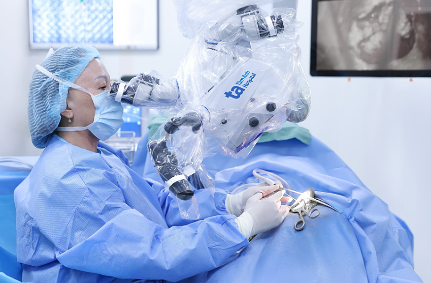 |
Prof. Chung Thuy performs cochlear implant surgery on a pediatric patient using a K.Zeiss Kinevo 900 surgical microscope. Illustration: Tam Anh General Hospital.
Before surgery, the patient's MRI and CT ear images are uploaded into the surgical microscope system. AI analyzes the cochlear shape and reconstructs a personalized 3D map directly on the screen. Surgeons can view this map in real-time and cross-reference it during the procedure, which reduces risks.
For young children, the surgical microscope is even more crucial. Their ear structures are smaller and narrower, making procedures more challenging than in adults. It is especially vital for pediatric patients with anatomical abnormalities.
You should research hospitals that use surgical microscopes for cochlear implant procedures to make an informed choice. Additionally, prioritize hospitals with experienced surgical teams to enhance effectiveness and minimize complications.
Prof. Dr. Tran Phan Chung Thuy
Director, Department of Otolaryngology
Tam Anh General Hospital TP HCM
| Readers can submit questions about ear, nose, and throat conditions here for a doctor's response. |