Phan Thi Hong Dang, a Master and Doctor from the Department of Radiation Therapy, Cancer Center, Tam Anh General Hospital TP HCM, stated that lung tumors constantly move with respiration. Consequently, even a minor deviation can cause radiation to affect the heart, healthy lung tissue, or esophagus. Currently, a combination of factors allows doctors to precisely target radiation.
Simulation CT scans
The initial step for precise lung tumor radiation therapy involves simulation CT scans, which create a three-dimensional image of the chest, tumor, and surrounding organs. Doctors may also order additional PET/CT or MRI scans to clarify tumor boundaries and assess the extent of invasion more accurately.
A key characteristic of lung tumors is their movement with respiration. Doctors utilize 4D simulation CT to record the tumor's path throughout the entire respiratory cycle. This allows for the precise identification of the treatment area closest to the tumor, preventing margin expansion and minimizing the radiation dose to healthy tissue.
Radiation dose planning
Based on simulation images, radiation oncologists and medical physicists calculate the beam direction, intensity, radiation delivery time, and safety margin, accounting for respiratory motion. This plan is individualized for each patient, ensuring the highest concentrated dose to the tumor while protecting the heart, healthy lung tissue, and surrounding organs.
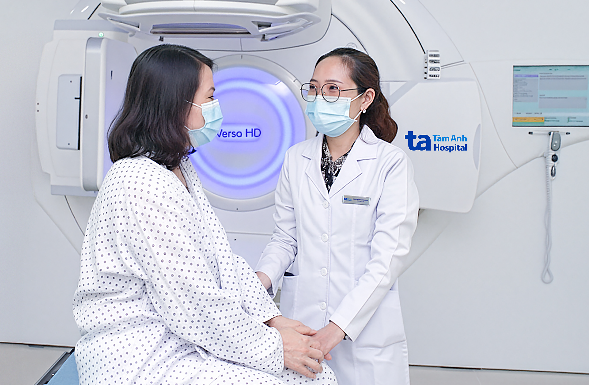 |
Doctor Dang encourages a patient before a radiation therapy session. *Illustration: Tam Anh General Hospital*
Respiratory rhythm control
Lung tumors can shift 1-2 cm during inhalation or exhalation. Consequently, patients may utilize one of two breathing techniques: deep inspiration breath hold (DIBH) or respiratory-gated radiation therapy.
With DIBH, patients take a deep breath and hold it for several seconds. As the chest expands, the distance between the tumor and vital organs (heart, healthy lung tissue) increases. This helps radiation precisely target the tumor and reduces the dose to healthy tissue. Conversely, with the respiratory-gated technique, the radiation machine only delivers radiation during the specific breathing phase when the tumor is within the target region (typically during exhalation). This ensures the radiation hits the tumor even when it is continuously moving.
Modern radiation therapy technology
According to Doctor Dang, modern radiation therapy techniques such as intensity-modulated radiation therapy (IMRT) and volumetric modulated arc therapy (VMAT) enable machines to adjust the beam shape and intensity precisely to the lung tumor's structure. The radiation is designed to conform closely to the tumor, delivering a high dose while minimizing the dose to healthy tissue.
Before each radiation therapy session, the image-guided radiation therapy (IGRT) system captures the patient's body position directly on the treatment couch. This new image is then compared with the simulation image. If there is a mismatch, the machine automatically corrects the deviation, which typically does not exceed 1 mm.
Achieving precise and safe lung tumor radiation therapy relies not only on technology and techniques but also on the crucial coordination among the team. This team includes radiation oncologists, medical physicists, and technicians. The radiation oncologist calculates the optimal dose and safety limits for each organ. The medical physicist verifies machine quality and the radiation therapy plan. Radiation therapy technicians secure the patient's position and monitor them throughout each session.
Nhat Minh
| Readers can submit questions about cancer here for doctors to answer. |