Mr. Phuong first tore his anterior cruciate ligament (ACL) in his right knee due to a sports injury, followed by a second tear from a traffic accident. According to Mai Hoang Duong, a Master of Science, Doctor, and First-Degree Specialist at the Orthopedic Trauma Center, Tam Anh General Hospital Ho Chi Minh City, recurrent ligament tears can lead to early joint degeneration, chronic pain, joint instability, muscle atrophy, and limited mobility.
To enable Mr. Phuong to resume sports activities and minimize the risk of future re-tears, doctors employed a combination of three techniques for the surgery: Lemaire, superficial quadriceps tendon, and All-inside.
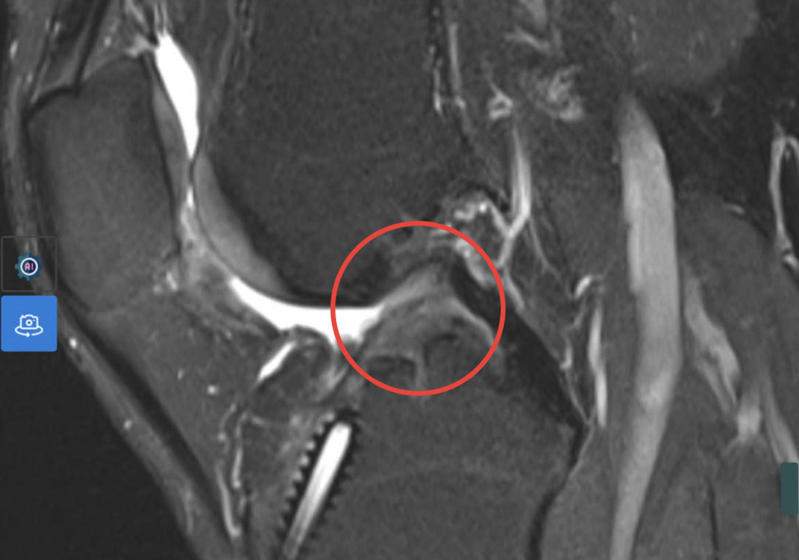 |
MRI results showed a complete anterior cruciate ligament tear. Photo: Tam Anh General Hospital.
Doctors conducted MRI and CT scans to reconstruct the knee joint. This allowed them to examine the existing tunnel and surrounding structures, guiding the selection of the tendon graft, fixation devices, and precise tunnel placement. The procedure began with an arthroscopy to directly assess intra-articular damage. Previous ligament fixation devices were removed, the area was cleaned, and a new tunnel was created using CT guidance. Subsequently, a segment of the superficial quadriceps tendon was harvested from the front of the thigh, above the patella, to form the ligament graft based on precise measurements.
Doctor Duong explained that this tendon is increasingly favored in ligament reconstruction. Its larger diameter provides a more substantial graft, enhancing stability and reducing the risk of re-tears. The tendon's optimal length also accommodates various fixation techniques, and its harvest site typically results in less pain and damage.
Following the ACL reconstruction with the superficial quadriceps tendon graft, doctors utilized the iliotibial band for external reinforcement via the Lemaire technique. This graft was threaded beneath the lateral ligament, tensioned, and secured to the bone. This approach mitigates rotational forces on the new graft, enhances knee joint stability, and reduces the likelihood of graft laxity and recurrent tears. Throughout the procedure, a thigh tourniquet was not used. This decision aimed to prevent post-operative pain, avoid nerve-muscle damage associated with tourniquets, reduce swelling and knee stiffness, lower the risk of venous thrombosis, and minimize fluid extravasation into soft tissues.
All intra-articular manipulations were performed through small 1 cm skin incisions. This advanced All-inside arthroscopic technique significantly minimizes damage to surrounding soft tissues, lowers the risk of infection, leads to less pain and blood loss, and facilitates a quicker recovery for the patient.
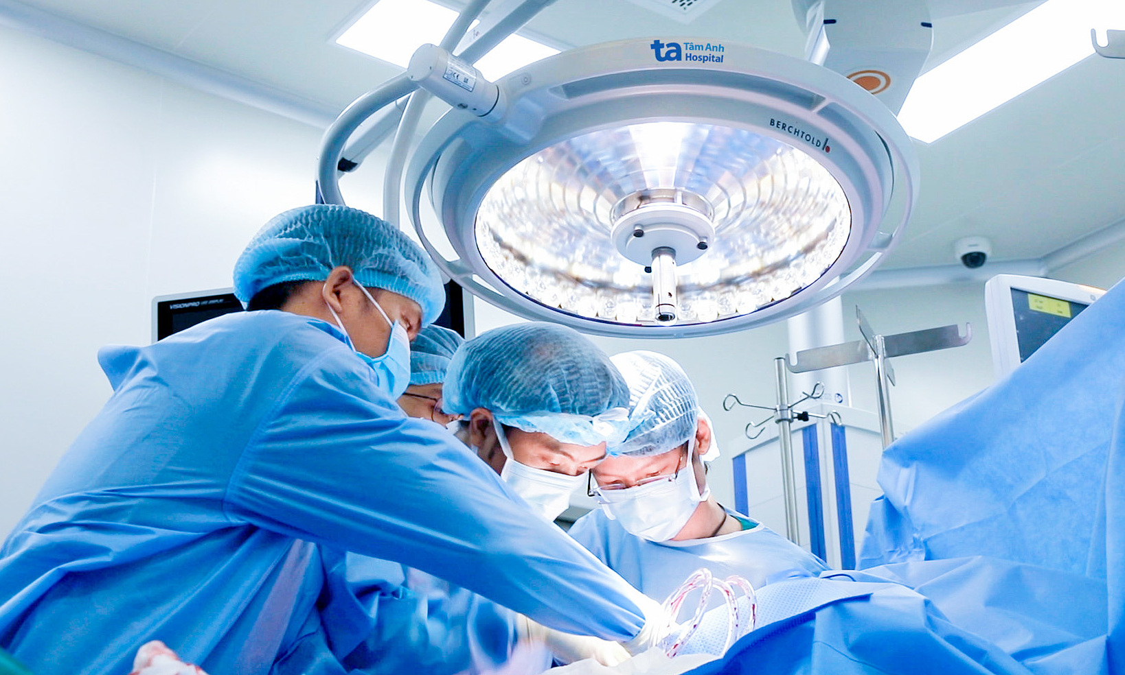 |
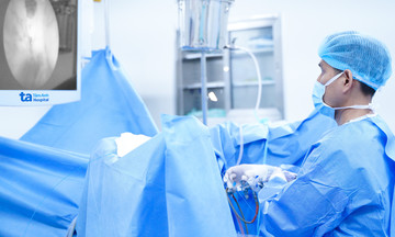
Doctor Duong (right) harvests the superficial quadriceps tendon for the patient. Photo: Tam Anh General Hospital.
On the first day post-surgery, Mr. Phuong reported only mild pain at the incision site and was able to walk with crutches. Doctors anticipate he will resume normal activities after 6 weeks and return to sports after 6 months.
Doctor Duong noted that recurrent ligament tears occur in approximately 3-10% of cases. Causes include new injuries at the original site, suboptimal surgical techniques, undersized grafts, or inadequate rehabilitation. Multiple ligament tears also lead to slower recovery rates in subsequent treatments and an elevated risk of re-tears. Therefore, individuals experiencing a first-time ligament tear should consult an orthopedic trauma or sports medicine specialist. Post-treatment, adhering strictly to a doctor-prescribed rehabilitation program is essential, and strenuous activities should be avoided prematurely. If any signs of a re-tear emerge, prompt medical consultation is advised for timely intervention.
Phi Hong
*Patient's name has been changed