A CT Somatom Force VB30 scan of Ba Lan at Tam Anh General Hospital, Hanoi, revealed a small ground-glass nodule, approximately 10 mm, classified as high risk. Signs of surrounding lung tissue retraction suggested an early-stage malignant lesion in the right middle lobe of the lung.
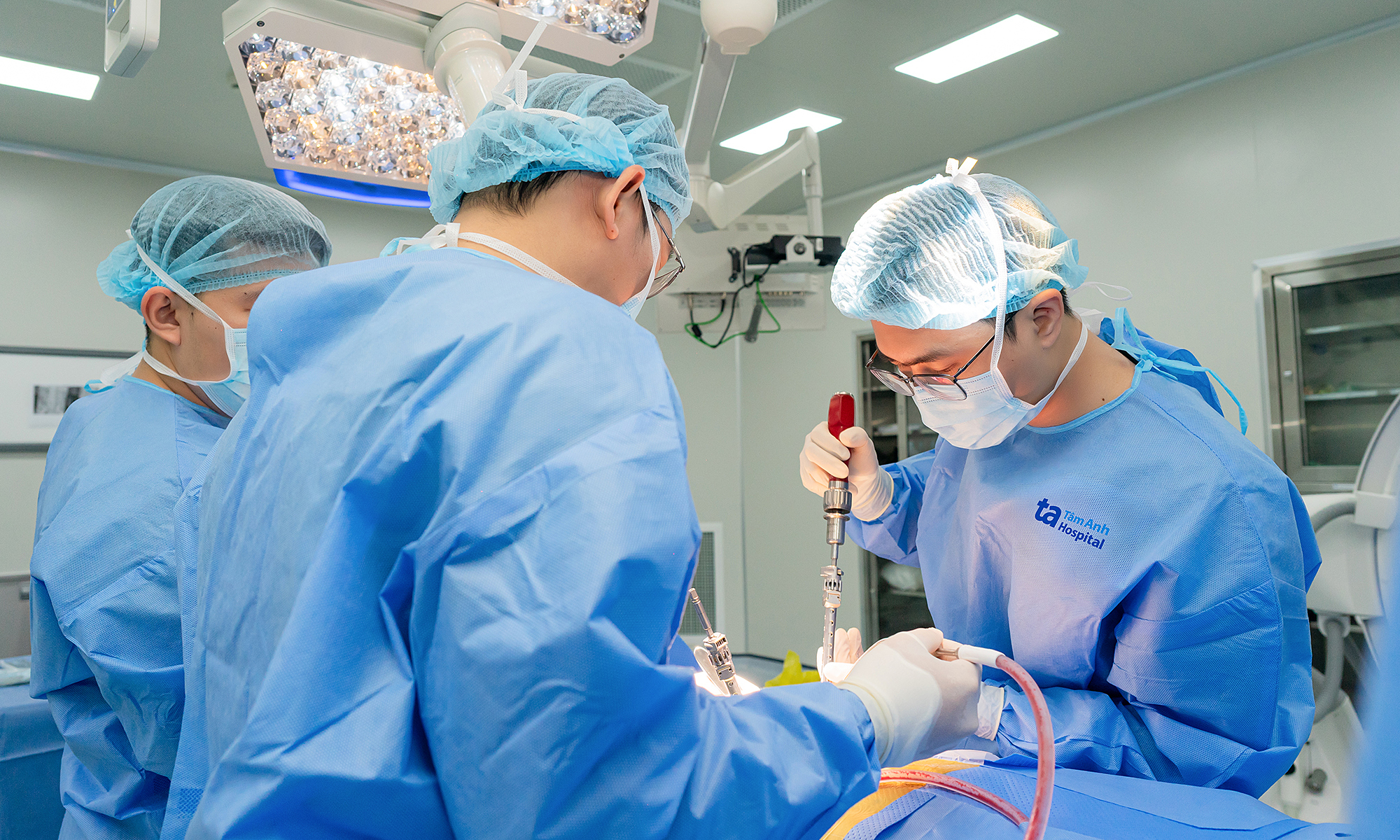
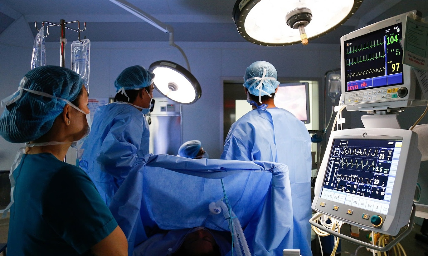
Associate Professor, Doctor, Specialist Level II Nguyen Huu Uoc, from the Department of Cardiovascular and Thoracic Surgery at Tam Anh General Hospital, Hanoi, ordered a lung nodule biopsy. This was performed using thoracoscopic wedge resection, a minimally invasive procedure that removes a small section of the lung containing the lesion. This method preserves healthy lung tissue if the lesion is not malignant.
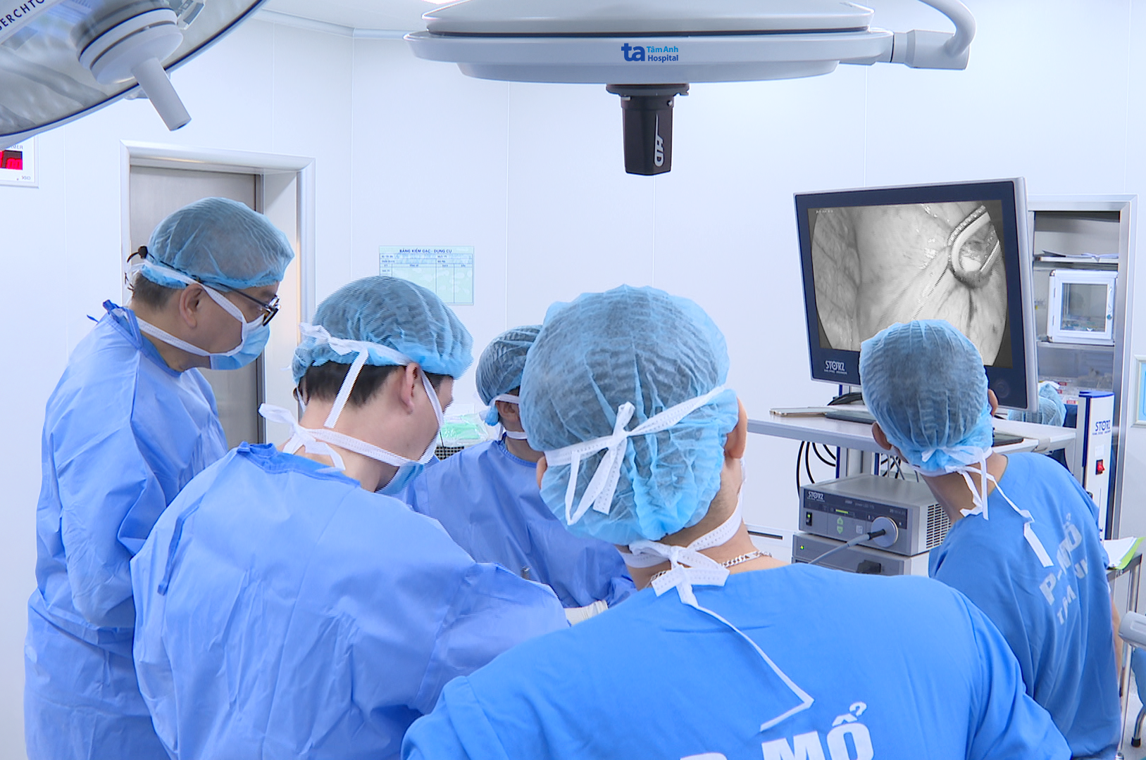 |
The surgical team accesses the lung parenchyma containing the lesion. Photo: Tam Anh General Hospital |
The tissue sample underwent immediate frozen section biopsy during surgery to determine the tumor's nature. Pathology results confirmed it was a malignant lesion. The surgical team then performed a right middle lobectomy, removing the tumor, combined with lymph node dissection.
Post-operative pathology confirmed adenocarcinoma of the lung, grade 2. The tumor measured only 1.1 cm in diameter, was confined to the lung parenchyma, and had not invaded blood vessels or nerves. According to the TNM (AJCC 8) classification, the case was categorized as stage pT1b, a very early stage of lung cancer.
Ba Lan recovered well and was discharged one week after post-operative monitoring. In addition to adhering to prescribed medication, she needs regular follow-up appointments and breathing exercises to restore lung function. Pathology and molecular biology test results will help doctors consider adjuvant treatments in the future, such as targeted therapy or immunotherapy. Patients should also undergo specialized tests like circulating tumor DNA (ctDNA) quantification and regular chest CT scans every 6-12 months to detect early recurrence risks.
According to Associate Professor Uoc, early-stage lung cancer often presents no symptoms. Manifestations such as persistent cough or mild fatigue are often non-specific and easily overlap with common respiratory illnesses. Many lung cancer cases are only incidentally discovered during routine health check-up chest CT scans.
Doctors recommend everyone undergo regular health check-ups. Individuals with risk factors, such as being over 30 years old, living in urban areas, frequent exposure to air pollution, or a family history of lung cancer, should consider low-dose chest CT screening every 12-24 months. Detecting lesions at an early stage increases the chance of radical treatment and reduces the burden of care.
Ly Nguyen
*Patient's name has been changed
| Readers can submit questions about cardiovascular health here for doctors to answer |