Electrocardiogram, Holter monitoring, and specialized tests at Tam Anh General Hospital Hanoi confirmed the patient had high-grade atrioventricular block, complete left bundle branch block, accompanied by heart failure and dyslipidemia. Mr. Son's coronary artery showed calcification and partial stenosis of approximately 50-60% in several segments. This level of damage was managed with medical treatment, not requiring stent placement.
Master of Science, Doctor Pham Van Thao, from the Cardiology Department, explained that high-grade atrioventricular block occurs when electrical signals from the atria do not transmit fully to the ventricles. This causes a slow and potentially unstable heart rate. Patients may experience fatigue, dizziness, hypotension, or syncope due to reduced blood flow to the brain and other organs. Severe cases can lead to heart failure or cardiac arrest.
The team implanted a dual-chamber permanent pacemaker using the left bundle branch area pacing (LBBAP) technique. This physiological pacing method involves inserting an electrode into the conduction system near the left bundle branch to stimulate the heart to contract in a manner close to its natural electrical activity.
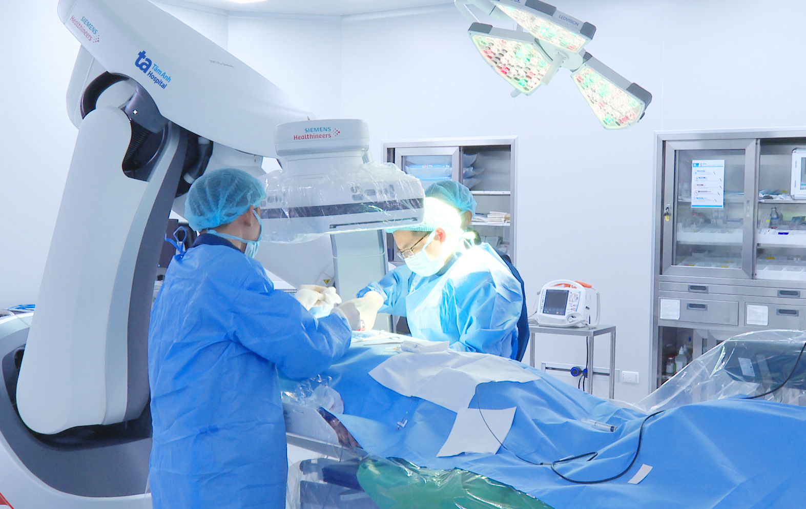 |
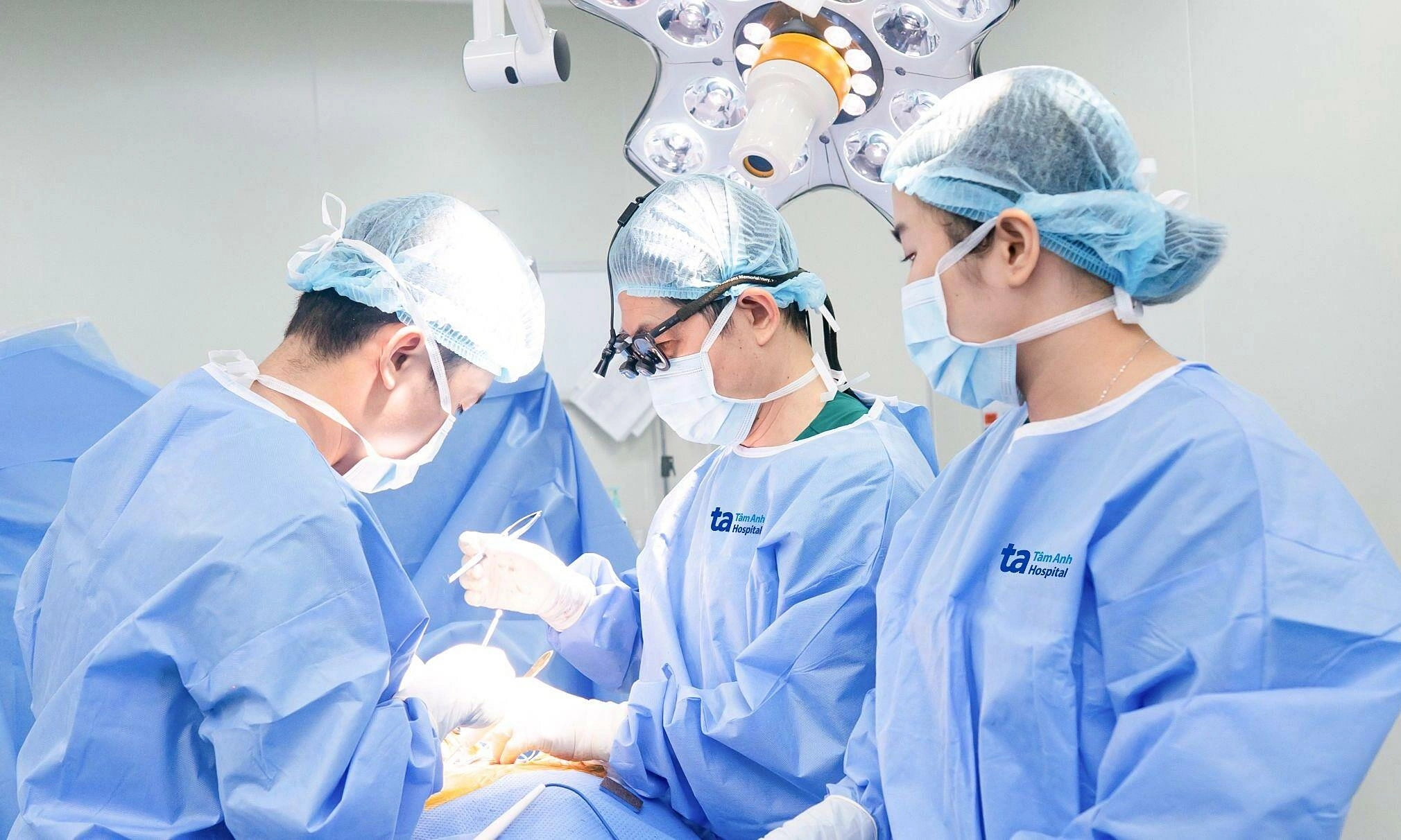
Doctor Thao and his team performing the LBBAP pacemaker implantation. *Photo: Tam Anh General Hospital* |
According to Doctor Thao, conventional right ventricular pacing, used previously, could cause ventricular dyssynchrony with high frequency during prolonged pacing, affecting heart function. LBBAP overcomes this limitation by directly stimulating the conduction system, helping the two ventricles contract more physiologically. For a 90-year-old patient, this method helps maintain a stable heart rate and reduces the impact of prolonged right ventricular pacing.
The team administered local anesthesia and inserted the electrode into the heart via a vein. Doctors checked electrical signals, stimulation thresholds, and electrocardiograms to confirm correct electrode placement in the left bundle branch area before connecting it to the dual-chamber pacemaker. The device was then placed under the skin in the chest area. The procedure was performed under the guidance of a digital subtraction angiography (DSA) machine and cardiac electrophysiology monitoring. After the intervention, Mr. Son's condition stabilized, his symptoms improved, and he was discharged.
Doctor Thao noted that the indication for this method primarily depends on the extent of the conduction disorder, symptoms, underlying diseases, general health, and the expected benefits of the intervention. For symptomatic high-grade atrioventricular block, maintaining an appropriate heart rate reduces the risk of syncope, heart failure, and dangerous events caused by bradycardia.
After pacemaker implantation, patients must take prescribed medications and attend follow-up appointments to check the device's function, electrode integrity, and adjust parameters as needed. In the initial weeks, patients should limit strenuous movement of the arm on the side where the pacemaker was implanted, as advised by the doctor. Patients should seek immediate medical attention if they experience severe fatigue, shortness of breath, dizziness, lightheadedness, or recurrent syncope; or if the implantation site becomes swollen, hot, red, increasingly painful, or discharges fluid.
When seeking medical care, patients must inform healthcare staff about their pacemaker. Magnetic resonance imaging (MRI) should only be performed if the pacemaker system is confirmed to be MRI-compatible and has been appropriately checked and programmed.
Thanh Ba
| Readers can submit questions about cardiovascular disease here for doctors to answer |