Ms. Tinh was being treated for type 2 diabetes and hypertension when she underwent a health check-up at the General Internal Medicine Department, Tam Anh General Hospital, Ho Chi Minh City. A low-dose computed tomography (ct) scan for cancer screening revealed a semi-solid lesion in the S2 segment of her right lung, measuring 15x17 mm. The lesion was close to and pulling on the pleura, classified as Lung-RADS 4A, with a malignancy risk of 5% to 15%. Lung-RADS is a classification system used to evaluate abnormal nodules on lung ct scans for cancer screening.
Before the lesion was discovered, Ms. Tinh had no symptoms such as cough, fever, or fatigue, and her underlying conditions were stable.
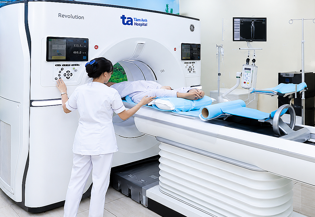 |
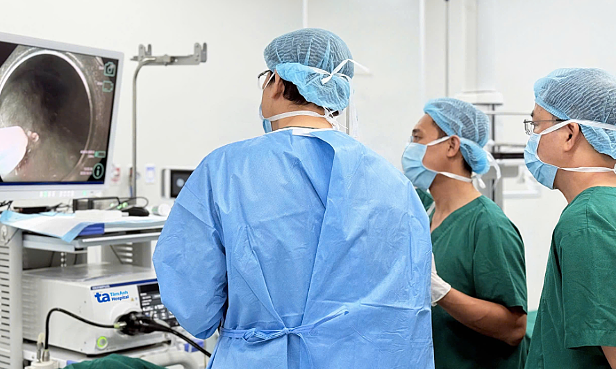
The patient underwent a low-dose lung scan using a 1975-slice ct machine, which detected the small lesion. *Photo: Tam Anh General Hospital*.
Doctor Ma Thanh Phong, Head of the Respiratory Unit, General Internal Medicine Department, stated that because the lesion was under 3 cm, it had not yet been definitively identified as a tumor. Doctors conducted tests for fungi and tuberculosis bacteria to determine the cause, and all results were negative.
The lesion was located in a hidden area, making it impossible to determine its nature through biopsy using flexible bronchoscope or transthoracic needle biopsy. Doctor Nguyen Anh Dung, Head of the Thoracic and Vascular Surgery Department, Thoracic and Vascular Surgery Center, prescribed laparoscopic surgery to address the lesion using the Da Vinci Xi robot, combined with intraoperative frozen section biopsy. If the lesion were benign, only tumor removal would be necessary; however, if it were malignant, a radical lobectomy would be performed to treat the cancer.
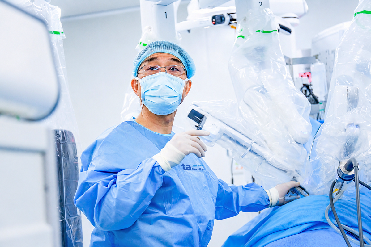 |
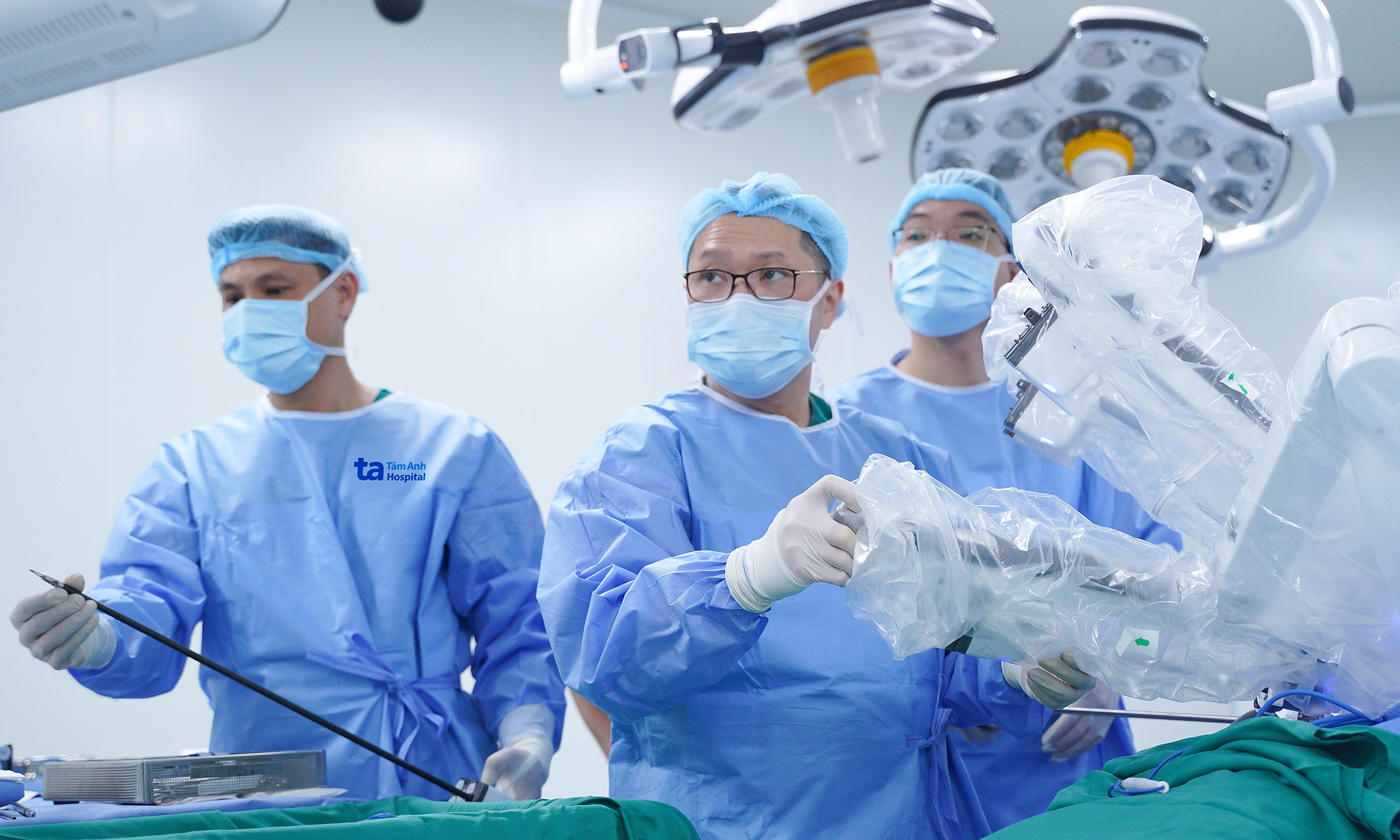
Doctor Dung connected the Da Vinci Xi robotic arm system to the patient's body before controlling the robot for surgery. *Photo: Tam Anh General Hospital*.
The flexible robotic arms navigated deep into narrow spaces within the chest cavity, helping the surgeon precisely locate the lesion and remove a portion of tissue containing the lung nodule in the upper lobe of the right lung for frozen section biopsy.
The tissue sample was immediately frozen at deep sub-zero temperatures (from -20 to -50 degrees Celsius), then cut into thin slices for microscopic examination. The results confirmed lung cancer, leading Doctor Dung to remove the upper right lung lobe containing the tumor.
The surgeon controlled the Da Vinci Xi robot to dissect and manage blood vessels layer by layer. This area features complex anatomical structures and a dense vascular network, demanding high precision. After one hour, the surgeon completed the upper right lung lobectomy and dissected the lymph nodes around the anatomical region.
 |
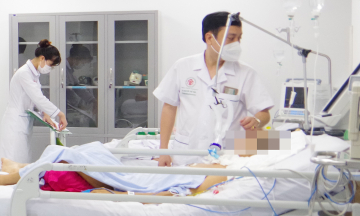
Doctor Dung checked on Ms. Tinh's health two days after surgery. *Photo: Tam Anh General Hospital*.
Two days after surgery, Ms. Tinh's health was stable, and she could move gently. Pathological results from the tissue sample confirmed stage one cancer, requiring no further treatment as it had been radically removed. The patient needs follow-up appointments as directed by her doctor.
Nhat Nhi
*Patient's name has been changed*
| From 17h30 to 20h30 on 19/5, Tam Anh General Hospital will host an online consultation titled "For a Healthy Vietnam - An Era of Growth, Applying Robotics and High Technology in Medical Examination and Treatment". The program will feature: Associate Professor Doctor Tran Quang Binh, Associate Professor Doctor Trieu Trieu Duong, Associate Professor Doctor Vu Le Chuyen, Associate Professor Doctor Nguyen Xuan Hien, and Doctor Pham Xuan Dung, all from the Tam Anh General Hospital system, the VNVC Vaccination Center system, and Eco Pharmaceutical Company. Readers can submit questions here for consultation. |