During a routine health check-up, Mrs. Hong, 67, discovered a 4 cm tumor in her right kidney. Initial advice suggested complete kidney removal to treat the cancer. However, Mrs. Hong sought an alternative at Tam Anh General Hospital, TP HCM, hoping for tumor excision and kidney preservation.
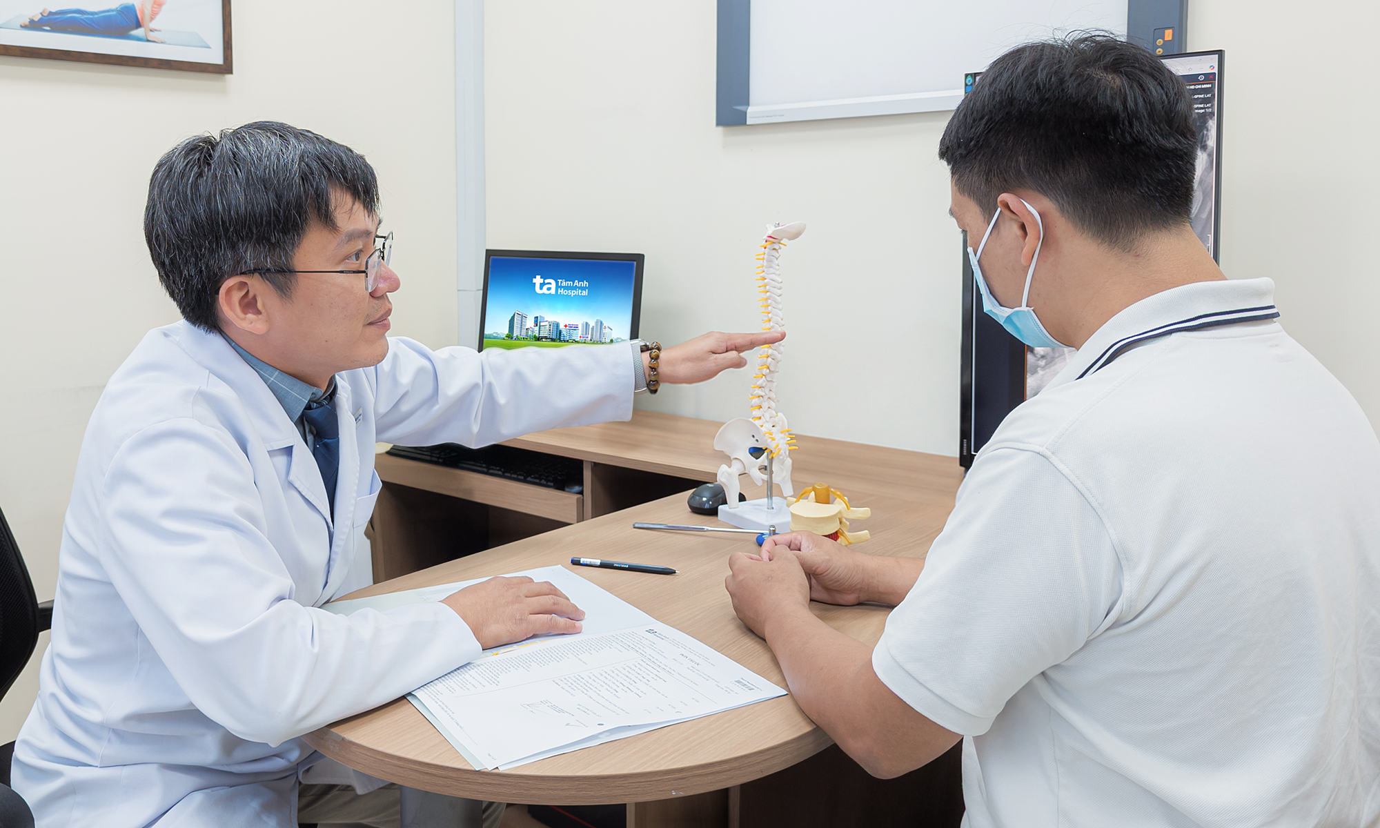
Dr. Le Phuc Lien, Head of the Female Urology Unit at the Center for Urology - Nephrology - Andrology, examined Mrs. Hong's case. He observed that the tumor was situated deep within the renal pelvis, a critical area containing major blood vessels and the kidney's urine collection system. "Removing the tumor is like defusing a bomb in a tangled web of wires", Dr. Lien stated. He added that during the procedure, even a one mm deviation could rupture a major blood vessel, leading to massive blood loss, or damage the entire renal pelvis system, rendering kidney preservation futile.
The surgical complexity was compounded by the kidney's limited tolerance to ischemia. According to Dr. Lien, a kidney can tolerate a maximum of 20-25 minutes of arterial clamping; exceeding this period causes cell death and irreversible functional loss. Within this narrow window, surgeons must meticulously excise the tumor, ensuring no cancer cells remain, and perform a complex three-layer suture, a task far more intricate than routine procedures. Inadequate suturing risks urine leakage into the abdominal cavity, potentially necessitating a second surgery to remove the entire kidney.
Given Mrs. Hong's age, removing one kidney would elevate her future risk of chronic kidney failure and cardiovascular diseases. Following a consultation, Dr. Lien opted for robotic kidney-preserving surgery, leveraging its precision to navigate these challenges.
Guided by a 3D screen magnified 10-15 times, the surgeon navigated robotic arms through the renal pelvis, parenchyma, and renal capsule. This allowed for precise dissection of the deep tumor while minimizing damage to healthy tissue. After tumor excision, a deep cavity connected to the urinary collection system was visible. The surgical team meticulously sutured the renal pelvis, achieved hemostasis in the renal parenchyma, and then reconstructed the outer capsule, nearly restoring the kidney's original structure.
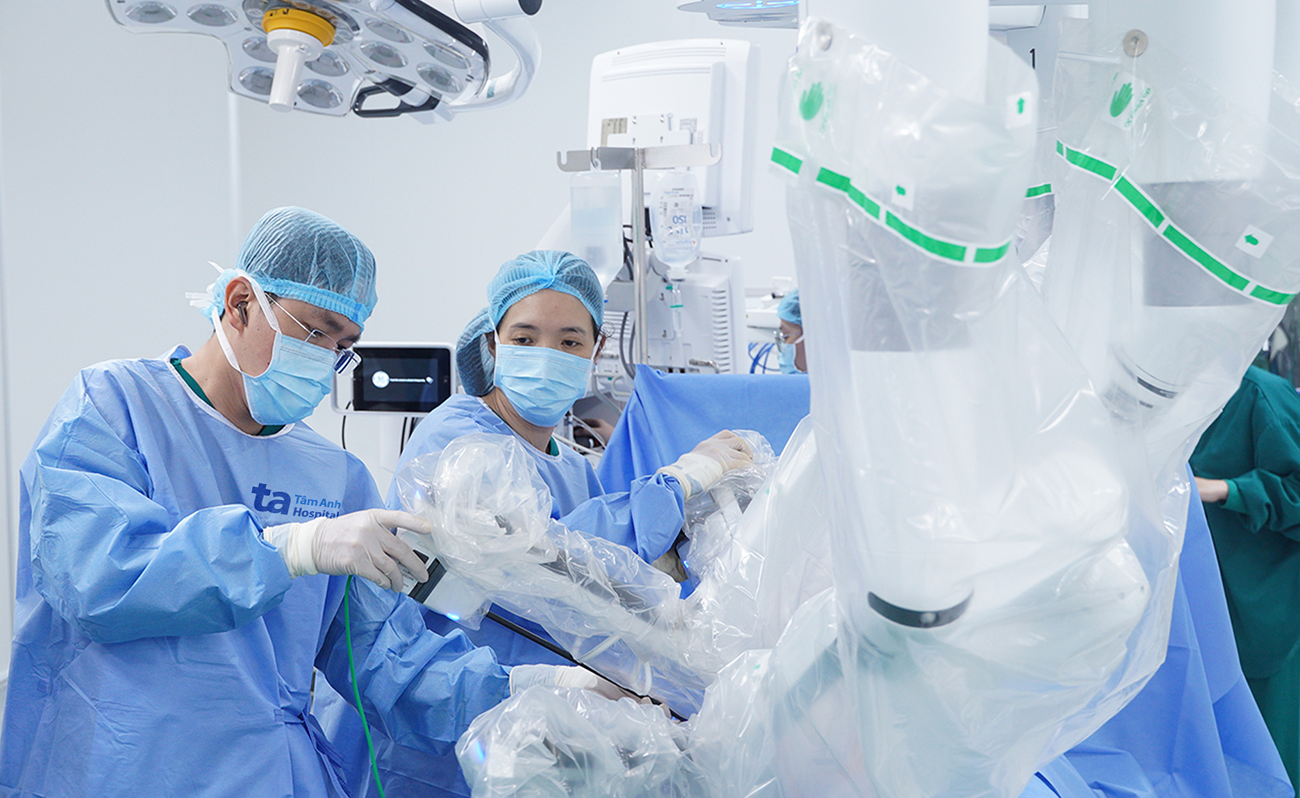 |
Dr. Le Phuc Lien (right) inserts the robotic arms into the patient's abdominal cavity in preparation for surgery. Photo: Tam Anh General Hospital |
By the 23rd minute, with the final sutures complete, Dr. Lien released the arterial clamp, and blood immediately perfused the kidney. The organ, initially pale, regained its healthy pink color, showing no signs of urine leakage or bleeding from the suture line. The tumor was then placed into a specialized bag and extracted through a trocar incision.
The entire surgical procedure, including anesthesia and robotic arm placement, took approximately two hours, significantly less than the over 3 hours typically required for similar complex cases. Within 24 hours, Mrs. Hong was able to sit up, drink water, and perform light movements, thanks to the small incision and minimal muscle pain. After three days, with stable kidney function and a dry incision, she was discharged.
Early-stage kidney cancer often develops asymptomatically, leading to incidental discovery during routine health check-ups. For localized tumors, the primary treatment objective is to preserve as much renal parenchyma as possible, rather than complete removal. This approach aims to maintain long-term kidney function and mitigate the risks of chronic kidney failure and cardiovascular complications. When tumors are large, invasive, or kidney preservation is unfeasible, surgeons may consider total nephrectomy, potentially combined with systemic therapies.
Individuals over 40 with risk factors or a family history of kidney cancer should undergo regular health check-ups to enable early detection, thereby improving the likelihood of kidney-preserving treatment.
Dinh Lam
*Patient's name has been changed.