79-year-old Mr. Ben lost vision in his left eye after an accident. Dr. Bui Viet Hung, Head of the Vitreoretinal Department at the High-Tech Eye Center, Tam Anh General Hospital in Hanoi, diagnosed him with complete conjunctival hemorrhage and a dense vitreous hemorrhage, obscuring the retina.
Mr. Ben's underlying atherosclerosis and use of anticoagulants presented significant challenges. Stopping the medication increased the risk of stroke due to his history of carotid artery stenosis, but continuing it raised the risk of prolonged bleeding during surgery. His acute bronchitis and laryngopharyngeal reflux further complicated the situation. After a hospital-wide consultation, the surgery was expedited to minimize the stroke risk.
The surgery presented a major challenge, as the extent of the injury was unclear. "We couldn't fully determine the damage pre-operatively and had to adapt intra-operatively," Dr. Hung said.
To ensure safety, the surgical team used specialized ophthalmic equipment and brain oxygen monitoring. Upon beginning the procedure, they discovered a large scleral tear causing the hemorrhages. They removed the damaged tissue and blood clots, then repaired the sclera.
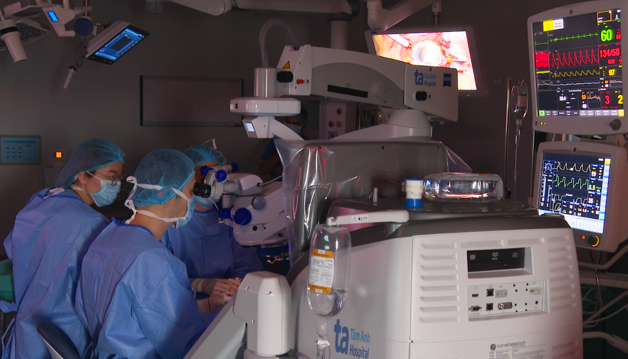 |
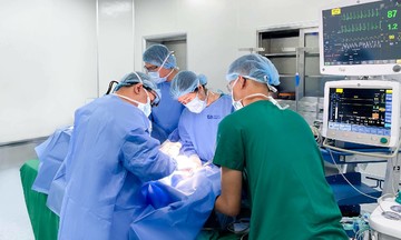
Operating in darkness to address a crushed eyeball and vitreous hemorrhage. Photo: Tam Anh General Hospital |
To enhance visibility, all operating room lights were extinguished, focusing the light solely on Mr. Ben's eye. The hemorrhaged vitreous was removed with a high-speed vitrectomy system. Examination revealed an undamaged retina, suggesting a positive prognosis. The 3-hour surgery was three times longer than anticipated.
 |
Mr. Ben recovered well after surgery and can walk unaided. Photo: Tam Anh General Hospital |
Mr. Ben was hospitalized for 9 days and discharged after his eye and overall health stabilized. Dr. Hung reports his vision has returned to normal.
Khue Lam
| Readers can submit questions about eye diseases here for doctors to answer. |