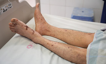
Ngoc, a 55-year-old woman with hypertension, type 2 diabetes, and dyslipidemia, recently experienced persistent pain in her left temple accompanied by progressively blurred vision. When the vision in her left eye significantly deteriorated and she developed electric shock-like numbness in her face, she sought medical attention at Tam Anh Hospital in TP HCM. An MRI 3 Tesla scan revealed two brain tumors. The larger tumor, measuring approximately 2.4 cm, was located on the left side of the cerebellopontine angle, deep within the internal auditory canal. It was compressing cranial nerves 5, 7, and 8, responsible for facial sensation, facial muscles, and hearing, respectively. The smaller tumor, measuring 1.8 cm, was situated in Meckel's cave, directly contacting the cavernous sinus segment of the internal carotid artery.
Dr. Chu Tan Si, Head of Neurosurgery at Tam Anh General Hospital's Neuroscience Center in TP HCM, described the case as complex due to the simultaneous presence of two tumors in the cerebellopontine angles on both sides of the brain, an area densely packed with cranial nerves and major blood vessels, and located near the brainstem. "This is a particularly sensitive surgical area, where even a few millimeters' deviation during the procedure could result in facial distortion, hearing loss, or permanent paralysis," Dr. Si explained, adding that without intervention, the tumors could obstruct cerebrospinal fluid flow, leading to brain herniation and ventricular dilation.
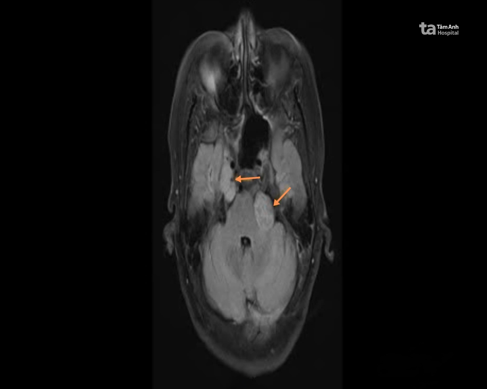 |
MRI image showing the two tumors near Ngoc's brainstem. Photo: Tam Anh General Hospital |
MRI image showing the two tumors near Ngoc's brainstem. Photo: Tam Anh General Hospital
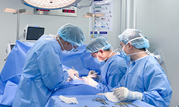
The surgical team decided to remove the larger tumor near the brainstem using the AI Modus V Synaptive robotic surgical system. Prior to the surgery, the robot's specialized AI software created a 3D model of the patient's brain structure. This allowed the surgeons to precisely determine the location, size, growth direction of the tumor, and its relationship to the cranial nerves and major blood vessels, enabling them to develop a detailed surgical plan.
During the surgery, the AI robot automatically adjusted the camera and lighting in real-time, maintaining a clear view in narrow areas like the internal auditory canal and Meckel's cave, where the tumor was close to nerves and blood vessels. The system also incorporated warning lights that activated when instruments approached critical nerves or blood vessels, assisting the surgeon in making precise movements.
The surgeon meticulously dissected the tumor layer by layer using microsurgical techniques, combined with a Cusa device. This device utilizes ultrasound waves to break down tumor tissue and aspirate it, removing the diseased tissue without damaging the surrounding healthy tissue. According to Dr. Si, this method is suitable for highly vascular tumors located deep within the brain, such as in Ngoc's case.
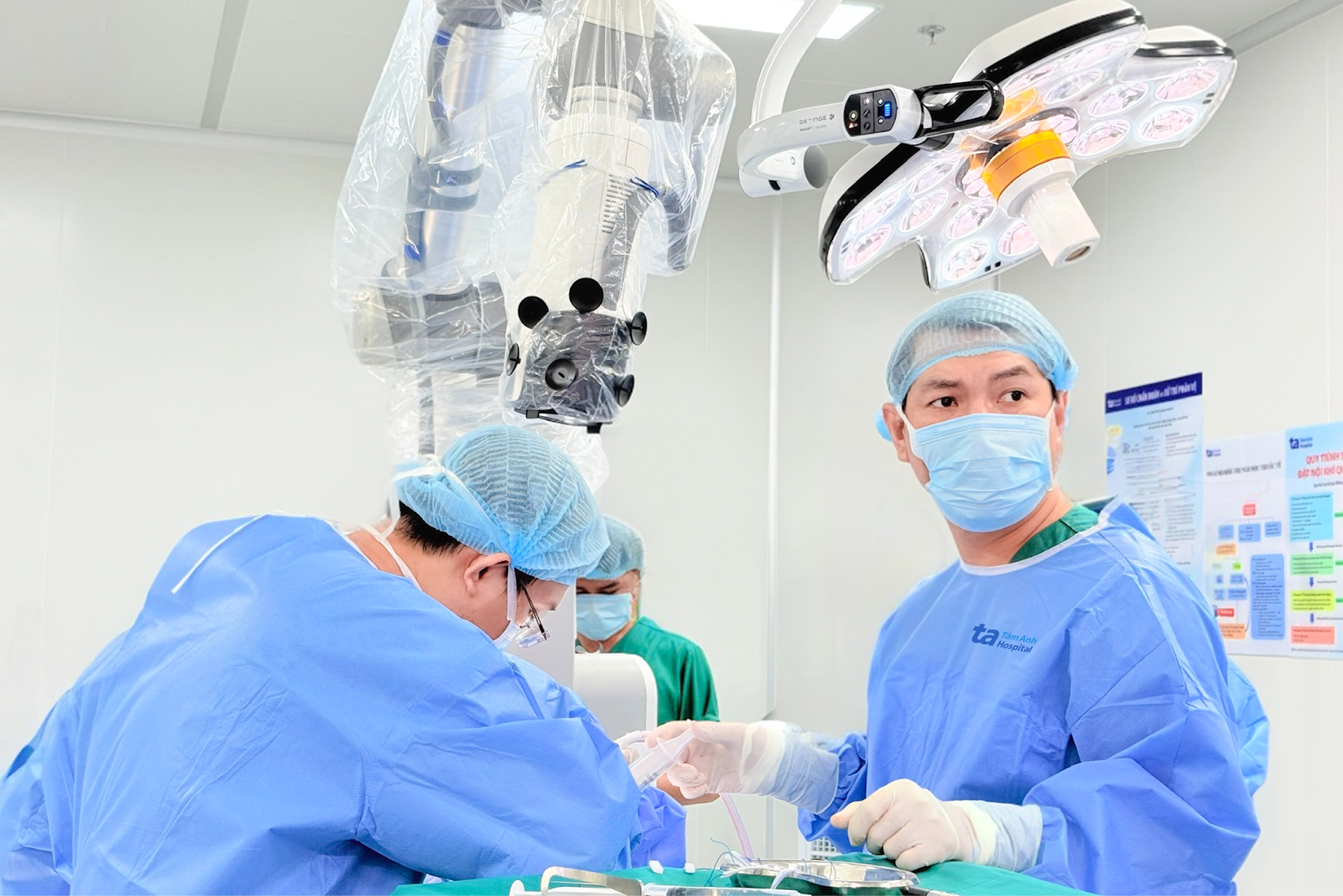 |
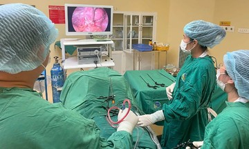
Surgeons using the AI robot and continuously monitoring the screen during Ngoc's brain tumor removal. Photo: Tam Anh General Hospital |
Surgeons using the AI robot and continuously monitoring the screen during Ngoc's brain tumor removal. Photo: Tam Anh General Hospital
After more than three hours of surgery, the team completely removed the larger tumor, preserving the facial motor nerves, hearing, and cranial blood vessels. The smaller tumor was mostly removed, with the remaining portion targeted for Gamma Knife radiosurgery. This non-invasive procedure uses focused gamma rays to destroy residual tumor tissue and is often employed for deep-seated, small, recurrent tumors, or those near high-risk areas like the brainstem or optic nerves. Post-operatively, Ngoc was alert, communicative, and experienced improved vision in her left eye, with no further facial numbness. On the third day after surgery, she could sit up, walk around her room, eat normally, and was discharged after a week.
The biopsy results confirmed an acoustic neuroma (schwannoma), a benign tumor that typically develops from the sheath of cranial nerves. These tumors can grow silently, compressing the brain and affecting vision, hearing, balance, and potentially causing serious complications.
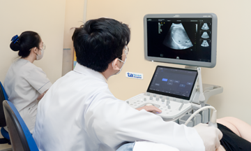
Doctors recommend that adults, especially those over 50 with chronic conditions, who experience persistent headaches, facial numbness, blurred vision, dizziness, or hearing loss, seek specialized medical evaluation. Advanced imaging techniques can detect tumors early, even before the onset of noticeable neurological symptoms.
Phuong Pham
*The patient's name has been changed.
| Readers can submit questions about neurological conditions here for doctors to answer. |