On 9/7, Dr. Chu Tan Si, Head of Neurosurgery at Tam Anh General Hospital's Neuroscience Center in Ho Chi Minh City, reported on the case of Yuta, a Japanese national. Yuta was admitted to the hospital suffering from prolonged fatigue, persistent headaches, and dizziness. While he hadn't experienced vomiting, he exhibited neurological signs suggesting a possible intracranial injury.
A 3 Tesla MRI scan revealed two subdural hematomas, one on each side of the brain. The left hematoma, larger at approximately 60 ml, was compressing surrounding brain tissue, narrowing the left ventricle, and shifting the midline of the brain about 1 cm to the right. The right hematoma, measuring around 30 ml, showed signs of left uncal herniation, indicating dangerous brain compression.
Yuta's diagnosis was chronic subdural hematoma resulting from a head injury that tore small veins, causing bleeding. According to Dr. Si, this condition often develops silently, with blood slowly leaking into the subdural space and accumulating over time, eventually compressing brain tissue. Without timely intervention, the growing hematoma can lead to decreased consciousness, hemiparesis, coma, and even death from brain herniation.
After consultation, doctors determined that bilateral burr hole surgery to drain the subdural hematomas was the optimal approach. This procedure allows for balanced decompression of both hemispheres, minimizing brain shift and damage to healthy tissue.
Using an AI-integrated neuronavigation system, the surgical team precisely located the burr hole sites, avoiding blood vessels and critical structures. They prioritized the left side due to the larger hematoma and higher risk of compression. Dark, clotted blood was evacuated, the cavity irrigated, and bleeding controlled with bipolar electrocautery. A drain was then inserted for continued post-operative drainage. The team then addressed the right hematoma, carefully monitoring intracranial pressure and ensuring proper cerebrospinal fluid circulation before placing a temporary drain to prevent recurrence. Both drains were connected to a closed system for sterility and close monitoring of post-operative drainage.
 |
Dr. Si and the surgical team perform the hematoma removal surgery on Yuto using AI-powered neuronavigation. Photo: *Tam Anh General Hospital* |
One day after surgery, Yuto was alert, pain-free, and had stable consciousness. His heart rate, blood pressure, and respiratory rates were all within normal limits. After three days, he could eat, walk normally, and was discharged after a week of observation, with follow-up appointments scheduled.
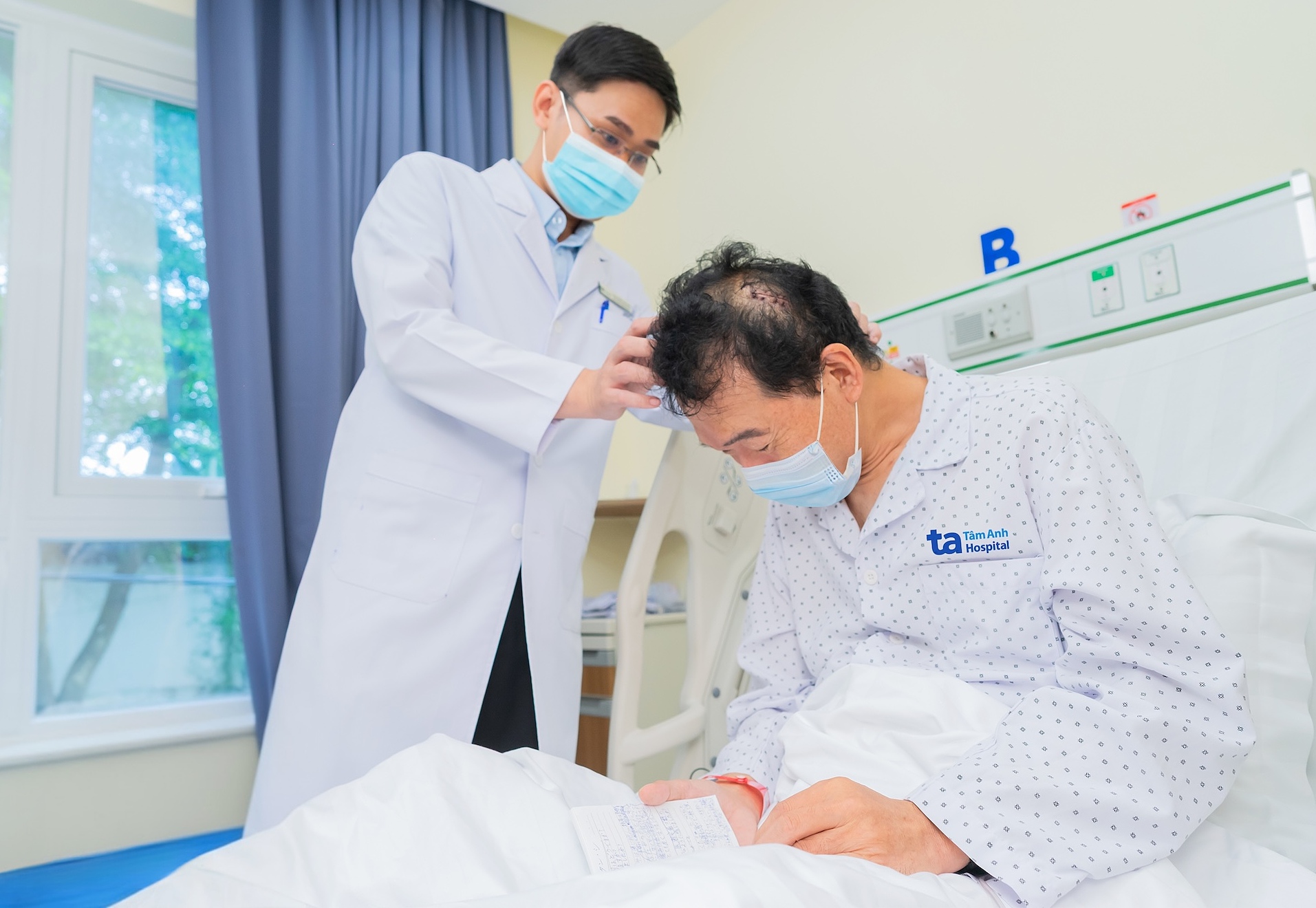 |
Dr. Si checks Yuto's incision post-surgery. Photo: *Tam Anh General Hospital* |
Chronic subdural hematoma is common in older adults, especially those with mild brain atrophy and fragile blood vessels. Even minor head trauma can tear bridging veins, causing slow, undetected bleeding that can lead to hematoma formation, according to Dr. Si. Symptoms often begin subtly, including headaches, fatigue, memory loss, mild motor impairment, or personality changes.
Dr. Si recommends that anyone who has experienced a head injury seek medical attention if symptoms persist. Depending on the individual's condition, diagnostic imaging, such as a 3 Tesla MRI, 768-slice CT, 1975-slice CT, or 100,000-slice CT scan, can help detect small, underlying brain injuries, enabling prompt intervention and preventing serious complications.
Lan Anh
*The patient's name has been changed.
| Readers can submit questions about neurological conditions here for doctors to answer. |