Ms. Dung sought treatment at Tam Anh General Hospital Ho Chi Minh City due to severe pain in her scalp and back of her neck, nasal congestion, and runny nose, for which medication provided no relief. She had previously undergone three surgeries for nasal septum deviation and chronic polysinusitis at another medical facility.
Professor Doctor Tran Phan Chung Thuy, Director of the Ear, Nose, and Throat Center, performed an endoscopy. He observed swollen nasal lining, opaque mucous discharge in the middle meatus, a perforated septum, and congestion. Computed tomography (CT) results revealed thickened lining in the maxillary and ethmoid sinuses on both sides, signs of previous maxillary sinus surgery, an S-shaped deviated nasal septum, and complete blockage of the frontal sinus by scar tissue.
Ms. Dung was scheduled for surgery. Given that all anatomical landmarks within the sinuses were lost after multiple previous operations, making it impossible to identify anatomical structures, the doctors decided to use an AI-integrated navigation system combined with endoscopy.
Before the operation, Ms. Dung's CT images were uploaded to the three-dimensional Navigation IGS system, which utilizes AI. The data was synchronized to create a 3D map of her nasal sinus anatomy, with surgical instruments displayed in real-time as the doctors operated. The team cleared the ethmoid sinus near the eye socket, then gradually drilled through the bone to access the frontal sinus, where a significant amount of pus was found.
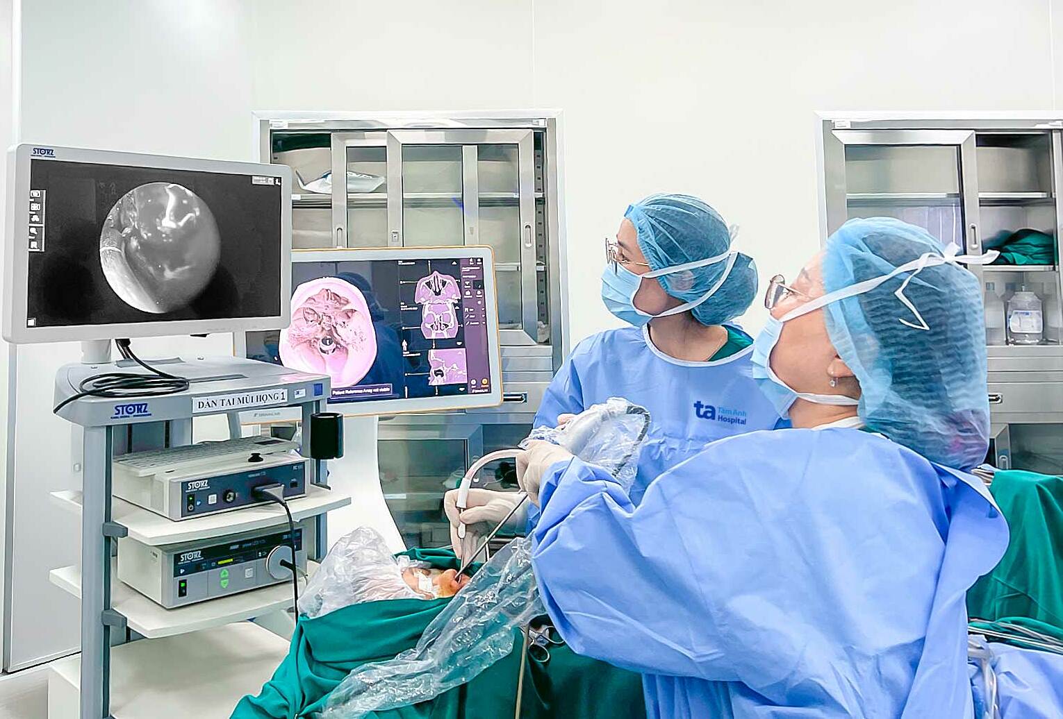 |
Professor Chung Thuy (right) observes the two navigation and endoscopy screens to find a safe approach to Ms. Dung's frontal sinus. Photo: Tam Anh Hospital.
Professor Chung Thuy explained that the frontal recess was blocked by scar tissue and fibrotic bone, preventing fluid drainage. This long-term fluid retention created an environment for bacteria to develop into an abscess, causing inflammation, increased pressure, and nerve irritation in the frontal region, leading to headaches. The team meticulously removed all inflamed tissue and pus. Finally, Professor Chung Thuy turned off the operating room lights and used a transillumination technique (where endoscopic light shines through the bone, skin, and inner corner of the eye) to inspect the frontal recess and the frontal sinus drainage pathway. He confirmed the frontal sinus was clear, concluding the surgery.
Following the surgery, Ms. Dung experienced no orbital or skull base complications. Her health recovered well, and she was discharged after two days. At her follow-up appointment, the surgical wound was healing properly, and her headaches and nasal congestion had significantly improved.
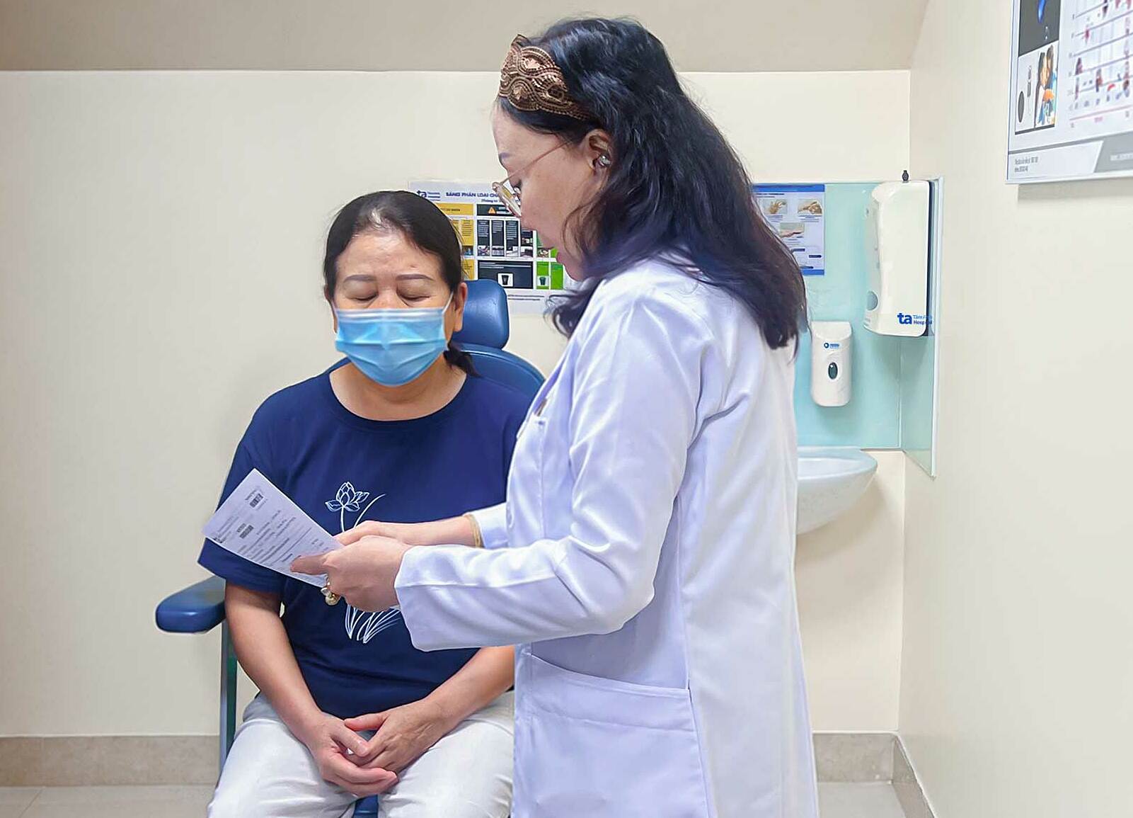 |
Ms. Dung at her 1-week post-op follow-up, with significant improvement in headaches. Photo: Hospital provided.
Professor Chung Thuy explained that the IGS navigation system creates a precise three-dimensional map simulating each patient's nasal sinus structure. Typically, recurrent polysinusitis surgery can combine navigation and endoscopy. However, Ms. Dung's case was particularly challenging because her frontal recess was already narrow and deep, further complicated by scar tissue, thick fibrotic bone, and a complete loss of anatomical structure. Therefore, combining IGS guidance, endoscopy, and transillumination was crucial to inspect the internal structures and enhance surgical safety.
Chronic sinusitis requires careful management to limit the risk of recurrence. After surgery, patients should regularly irrigate their nasal sinuses with saline solution, wear face masks, avoid smoke, dust, and cold air, and attend scheduled follow-up appointments for early detection and treatment of any abnormalities.
Uyen Trinh
| Readers can submit questions about ear, nose, and throat conditions here for doctors to answer |