Mr. Hung, 56 years old, had undergone four rounds of chemotherapy at a hospital in Hanoi before continuing his treatment at Tam Anh General Hospital Ho Chi Minh City. A CT scan revealed a tumor in the anterior mediastinal (chest) region, measuring 114x92x90 mm, showing signs of cardiac compression and approximately 7 mm of fluid accumulation.
Associate Professor, Doctor Vu Huu Vinh, Director of the Thoracic - Vascular Surgery Center, stated that the tumor was located within the pericardium, adhering to and deeply invading the atria, ventricles, and large surrounding blood vessels. The tumor occupied nearly half of the heart's volume, compressing it and leading to heart failure and acute cardiac tamponade due to the gradual increase in pericardial fluid, which obstructed blood flow back to the heart and circulation to the body.
Following a consultation, the surgical team decided to perform Da Vinci Xi robotic endoscopic surgery to minimize invasiveness and reduce trauma. Without robotic assistance, the team would have faced a high-risk open surgery involving sternal sawing.
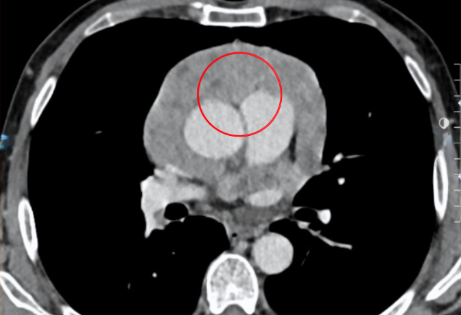 |
CT image showing the thymoma (red circle) compressing the patient's heart. Photo: Tam Anh General Hospital |
CT image showing the thymoma (red circle) compressing the patient's heart. Photo: Tam Anh General Hospital
Leveraging the advantages of a high-definition 3D camera, providing 10-15 times magnification, and flexible robotic arms, the team clearly visualized every small blood vessel and the micro-adhesion boundaries. Associate Professor Vinh controlled the robot, meticulously dissecting tightly adhered fibrous tissue layers, separating the large tumor from the atrial and ventricular surfaces, and completely freeing the vascular system. Subsequently, the team completely removed the tumor and excised the portion of the pericardium invaded by the tumor to fully decompress the heart.
Post-operative pathology results confirmed the lesion as a thymoma, mixed type B1 90% and B2 10%, with low malignancy. The patient did not require adjuvant treatment after surgery.
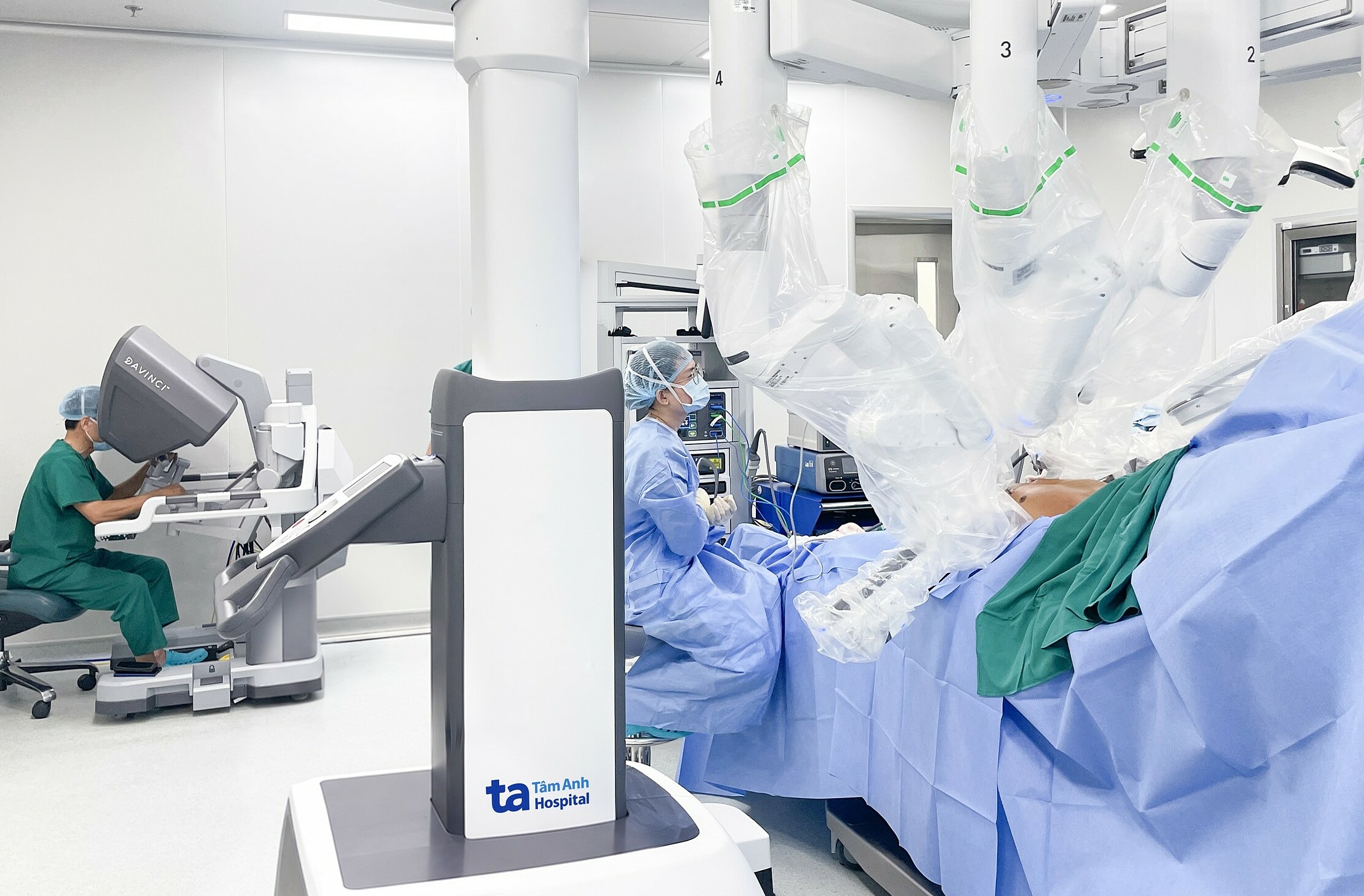 |
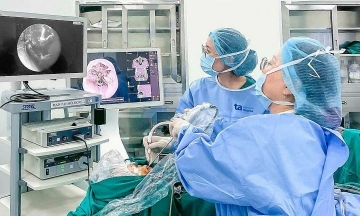
Associate Professor Vinh (left) controls the surgical robot to remove the chest tumor from the patient. Photo: Tam Anh General Hospital |
Associate Professor Vinh (left) controls the surgical robot to remove the chest tumor from the patient. Photo: Tam Anh General Hospital
Post-surgery, Mr. Hung's health progressed well with no complications. His symptoms of chest tightness and shortness of breath resolved, and his incision pain was minimal. He was discharged after three days.
Thymomas develop from thymic epithelial cells and often progress silently. Most patients do not experience symptoms or noticeable signs in the early stages. By the time symptoms become apparent, the tumor has usually advanced to a late stage, growing large enough to cause compression and deep invasion of surrounding organs.
Patients experiencing persistent chest pain or tightness, shortness of breath, a chronic cough, or unusual swallowing difficulties should seek medical examination. If a chest tumor has been detected, regular follow-up appointments and adherence to prescribed treatment are crucial to prevent tumor growth and complications.
Bao Anh
* Patient's name has been changed
| Readers can submit questions about cancer here for doctors to answer |