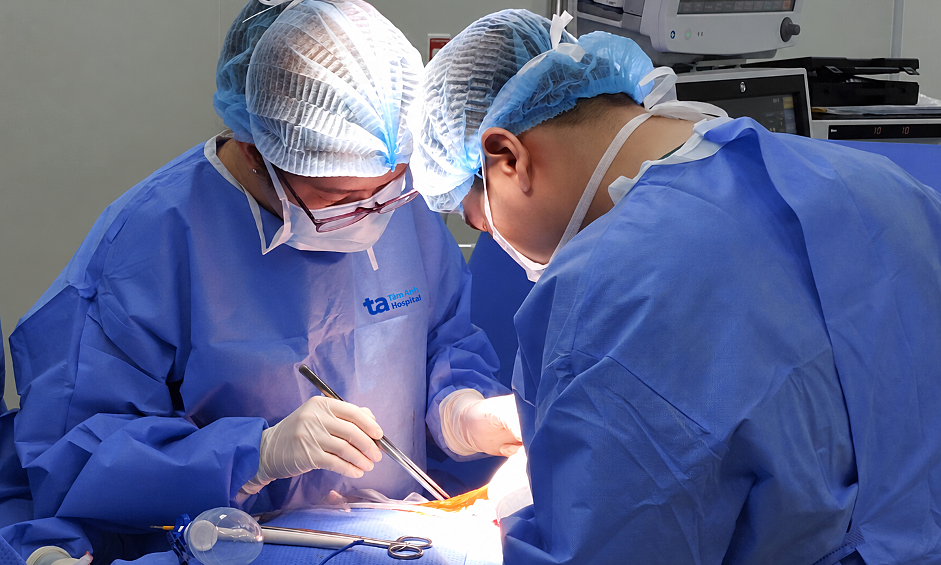
Mr. Tien's symptoms began one day before admission. Initially, he attributed them to fatigue and a vestibular disorder, resting and taking painkillers. The following day, he experienced vomiting, extreme exhaustion, and weakness and numbness on his left side, prompting his transfer to Tam Anh General Hospital, Ho Chi Minh City.
Doctor Pho Thien Phuoc, a Level I Specialist in the Emergency Department, reported that Mr. Tien was admitted with continuous vomiting, pupils dilated to approximately 5 mm on both sides, poor light reflex, right eyelid ptosis, facial nerve paralysis, and muscle strength of 3/5 on the left side, raising suspicion of acute brain injury.
CT scan results revealed blood in multiple brain areas, including the perimesencephalic cisterns, around the right cerebrum, and within the subarachnoid space of the right temporal region. The right occipital lobe brain tissue showed signs of ischemia, alongside a right posterior cerebral artery aneurysm, P1-P2 segment, measuring approximately 6x5 mm, with an irregular neck measuring about 5 mm.
Doctors diagnosed the patient with an intracranial hemorrhage caused by a ruptured posterior cerebral artery aneurysm. A brain aneurysm occurs when a blood vessel wall weakens and bulges out like a small sac. When an aneurysm ruptures, blood leaks into the subarachnoid space or other intracranial areas, potentially causing sudden severe headache, vomiting, altered consciousness, or focal neurological deficits.
Following a consultation, doctors decided to perform cerebral angiography and intervention using digital subtraction angiography (DSA) instead of open craniotomy to access the aneurysm. This approach aimed to reduce the risk of damage to brain tissue and adjacent nerves for the patient.
 |
The intervention team occluding the bleeding aneurysm for Mr. Tien. *Photo: Tam Anh General Hospital* |
With the assistance of the DSA system, Doctor Duong Dinh Hoan, a Level II Specialist and Head of the Neurointervention Unit at the Center for Diagnostic Imaging and Interventional Radiology, along with his team, inserted a catheter from the femoral artery to the left vertebral artery to examine the posterior cerebral circulation system.
After confirming the rupture of the right posterior cerebral artery aneurysm, doctors proceeded to carefully navigate a microcatheter into the aneurysm. They then sequentially deployed metal coils inside to occlude it. Post-intervention imaging confirmed the aneurysm was completely sealed, with no blood flow into the sac.
One day after the intervention, Mr. Tien was able to eat and resume normal activities. He was discharged two days later.
Patients experiencing sudden, severe headaches, especially when accompanied by dizziness, nausea, or weakness or numbness on one side of the body, should seek medical attention promptly. Early diagnosis allows doctors to identify the cause and initiate timely treatment. In cases of a ruptured brain aneurysm, prompt diagnosis enables rapid control of the bleeding source, reducing the risk of re-hemorrhage and more severe brain damage.
Nhu Ngoc
*Patient's name has been changed
| Readers can submit questions about neurological conditions here for doctors to answer |