X-ray and computed tomography (CT) scans both utilize X-rays (ionizing radiation) to generate images of the body's interior. The radiation dose is tightly controlled at relatively low levels. A single chest X-ray exposes the body to approximately 0,1 mSv, equivalent to the natural background radiation absorbed over about 10 days. A chest CT scan, however, involves a higher radiation dose, averaging around 6 mSv, depending on the equipment and technique used.
Radiation from these imaging techniques does not cause immediate poisoning or damage. Any potential effects are primarily long-term risks with a very low probability. In other words, undergoing an X-ray or CT scan does not mean a patient will develop cancer in the future.
According to the US National Cancer Institute (NCI) and the US Food and Drug Administration (FDA), the risk of developing cancer linked to a single diagnostic CT scan is very low. In most cases, the benefits of accurate disease detection and timely treatment outweigh this minimal risk.
To ensure patient safety, CT scan indications always follow two international principles. The first is to perform the scan only when truly necessary, when the results can aid in diagnosis or alter the course of treatment. The second is to optimize the radiation dose according to the ALARA (As Low As Reasonably Achievable) principle, meaning the radiation dose is reduced to the lowest possible level while still maintaining image quality for diagnostic purposes.
Many new generation CT systems significantly reduce patient radiation exposure. For instance, the Somatom Force VB30 CT system uses dual-source technology with very fast scanning speeds, allowing for a full-body scan in about 2 seconds. Its Admire image reconstruction technology can reduce X-ray doses by up to 85% compared to many previous CT generations, while still ensuring image quality.
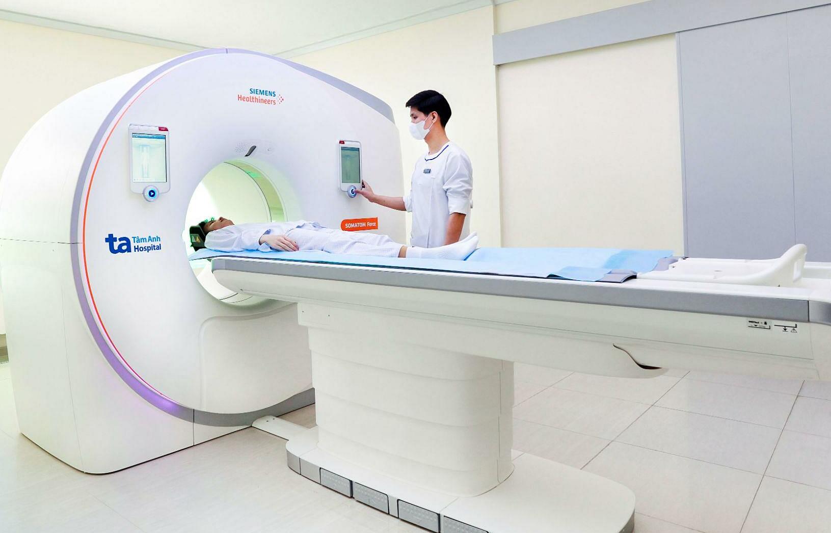 |
A patient undergoing a lung CT scan using the Somatom Force VB30 CT system. Illustration: Tam Anh Cau Giay General Clinic
Your doctor's decision to order both an X-ray and a CT scan of the lungs on the same day likely stems from the need to assess your condition at different levels. An X-ray serves as an initial imaging method, helping to detect relatively clear abnormalities in the lungs and chest through two-dimensional images. However, small lung nodules, lesions deep within the lung parenchyma, apical regions, or the mediastinum may not be clearly visible on an X-ray film. Additionally, X-ray images do not provide complete information about the internal structure of a lesion.
When a more detailed evaluation of the lung parenchyma, airways, or pleura is required, or to precisely determine the location, size, and characteristics of a lesion through three-dimensional cross-sectional images, doctors prescribe a chest CT scan. Therefore, X-ray and CT are not interchangeable techniques but rather complementary, helping doctors assess a patient's condition comprehensively.
A follow-up X-ray after about 20 days of treatment is typically performed to evaluate the treatment response, monitor how lung lesions are improving, and check for any complications. This is a common indication for patients with pneumonia, pulmonary tuberculosis, persistent lung lesions, or other respiratory diseases requiring progression monitoring.
The doctor's indications for your X-ray and lung CT scans are based on clear professional grounds. You should keep all imaging results to allow your doctor to compare them during follow-up visits. You should not arbitrarily refuse or delay follow-up scans when professionally indicated.
Dr. Nguyen Xuan Phong
Diagnostic Imaging Unit
Tam Anh Cau Giay General Clinic
| Readers can send respiratory questions here for a doctor's answer. |