Doctor Nguyen Trung Duc, a first-degree specialist at the Imaging Diagnosis and Interventional Radiology Center, Tam Anh TP HCM General Hospital, suspected Anh Khanh's sudden and escalating headaches indicated a brain injury. He ordered a 3 Tesla MRI scan. The results revealed a vascular malformation, approximately 5.5 x 4 x 3 cm in size, resembling a chicken egg, located in the right frontal lobe. The lesion consisted of numerous abnormal, intertwined blood vessels forming a nidus – a tangled mass connecting feeding arteries and draining veins.
Within this malformed vascular cluster, multiple microhemorrhages were present, possibly indicating previous bleeding. The images showed the malformation received blood from branches of the right middle cerebral artery and drained through a system of dilated cerebral veins. Doctors diagnosed Anh Khanh with an arteriovenous malformation (AVM) in his right frontal lobe.
Doctor Duong Dinh Hoan, a second-degree specialist and Head of the Neuro-Intervention Unit at the Imaging Diagnosis and Interventional Radiology Center, explained that in normal cerebral circulation, blood flows from arteries through a capillary network to supply oxygen and nutrients to brain tissue before returning to the venous system. In individuals with a brain arteriovenous malformation, arteries and veins connect directly via an abnormal vascular system, causing some blood to bypass the normal capillary network and flow straight from arteries to veins.
This direct flow of high-pressure arterial blood into the venous system can create abnormal pressure on vessel walls. When vessels within an AVM rupture, patients face a risk of cerebral hemorrhage, which can lead to severe headaches, weakness or paralysis, and altered consciousness.
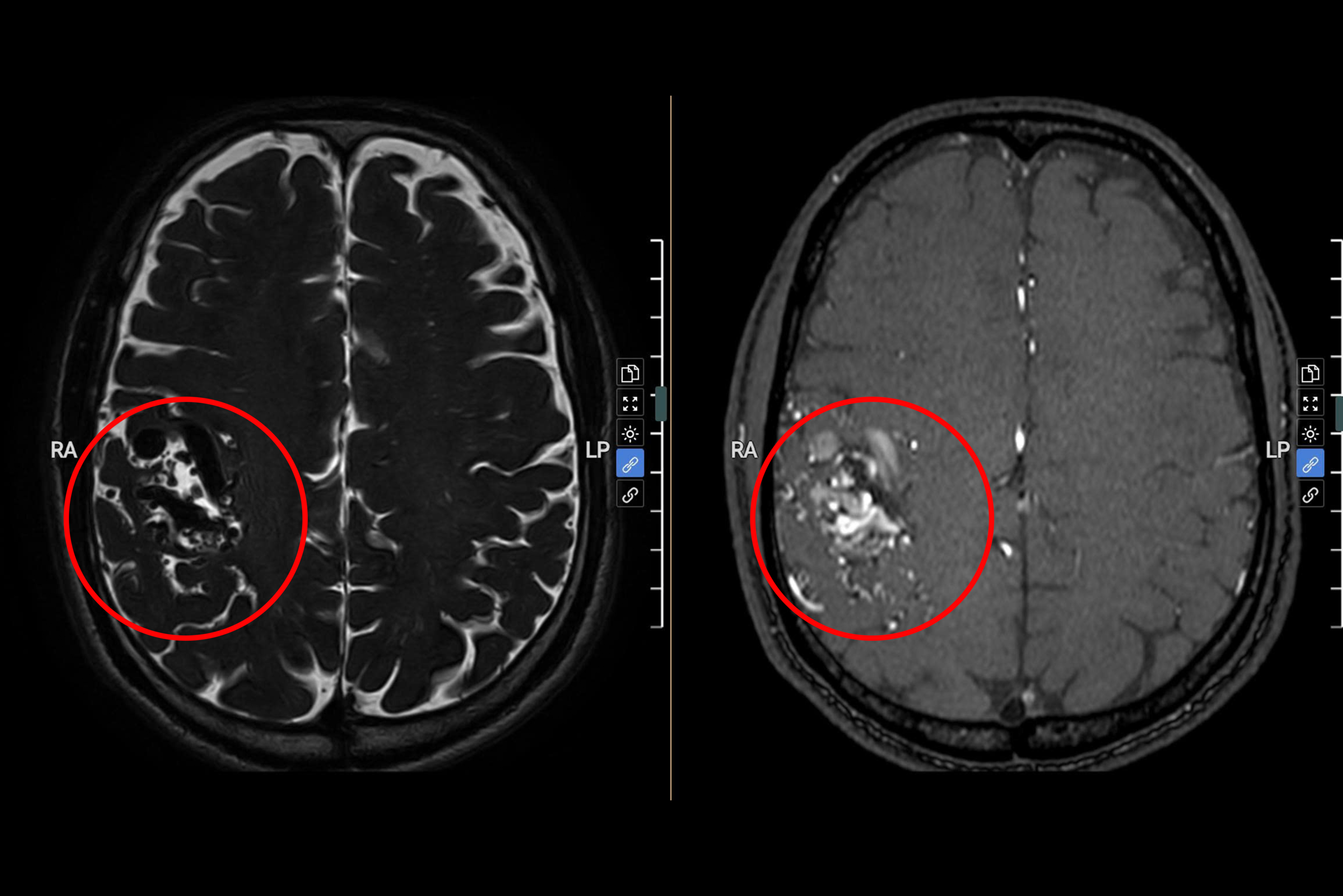 |
MRI images show a large vascular malformation in the frontal lobe. Photo: Tam Anh General Hospital |
Following a consultation, doctors decided to perform endovascular intervention guided by a digital subtraction angiography (DSA) system.
Anh Khanh underwent a DSA scan to detail the vascular structure before the intervention. The results showed the malformation was primarily in the right frontal lobe, receiving blood from branches of the right middle cerebral artery and draining through cortical veins into the superior sagittal sinus.
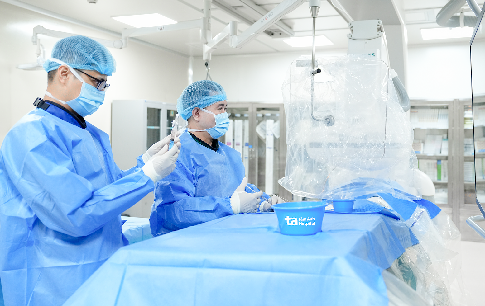 |
The medical team performed endovascular intervention for Anh Khanh under the guidance of a digital subtraction angiography (DSA) system. Photo: Hospital provided |
Doctors inserted a microcatheter from the right femoral artery up to the right internal carotid artery, selectively accessing a middle cerebral artery branch supplying the malformation. Through the microcatheter, embolization material was delivered to block the abnormal connection between the artery and vein. Post-intervention DSA images confirmed the targeted connection was occluded, and blood flow into the malformation significantly reduced.
One day after the intervention, Anh Khanh's headache subsided, with no neurological abnormalities recorded. He was able to walk independently and was discharged on the second day.
Brain arteriovenous malformations can exist for extended periods without obvious symptoms. Doctors advise that if new, unusual, or escalating headaches occur, patients should seek medical attention for examination and diagnosis. Depending on the condition, doctors select appropriate treatment methods such as surgery, radiosurgery, endovascular intervention, or a combination of approaches.
Nhu Ngoc
*Patient's name has been changed