According to Doctor CKI Duong Pham Van Thanh of the Emergency Department at Tam Anh General Hospital, Ho Chi Minh City, the patient's right upper lung lobe showed an area of consolidation approximately 37x25x27 mm. Within the lesion, an area of liquefactive necrosis had formed a cavity containing air, with thickened bronchial walls and fluid retention.
Mr. Di's lung images resembled a "tree-in-bud" pattern. This type of lesion is often associated with small bronchioles and airways, indicating a lung infection.
 |
Doctor Thanh examines Mr. Di. Photo: Tam Anh General Hospital |
Doctor Thanh explained that when a lung region sustains prolonged injury, its vascular system often changes to increase blood flow to the damaged area. Bronchial arteries can dilate, proliferate, and form abnormal branches.
These tortuous vessels have thin walls and are prone to injury, potentially becoming a source of bleeding into the airway. Normally, the lungs receive blood from two main systems: the pulmonary artery delivers blood for gas exchange, while the bronchial arteries supply oxygen-rich blood to the bronchi and a portion of the lung tissue.
 |
Abnormal proliferating blood vessels in Mr. Di's lung. Photo: Tam Anh General Hospital |
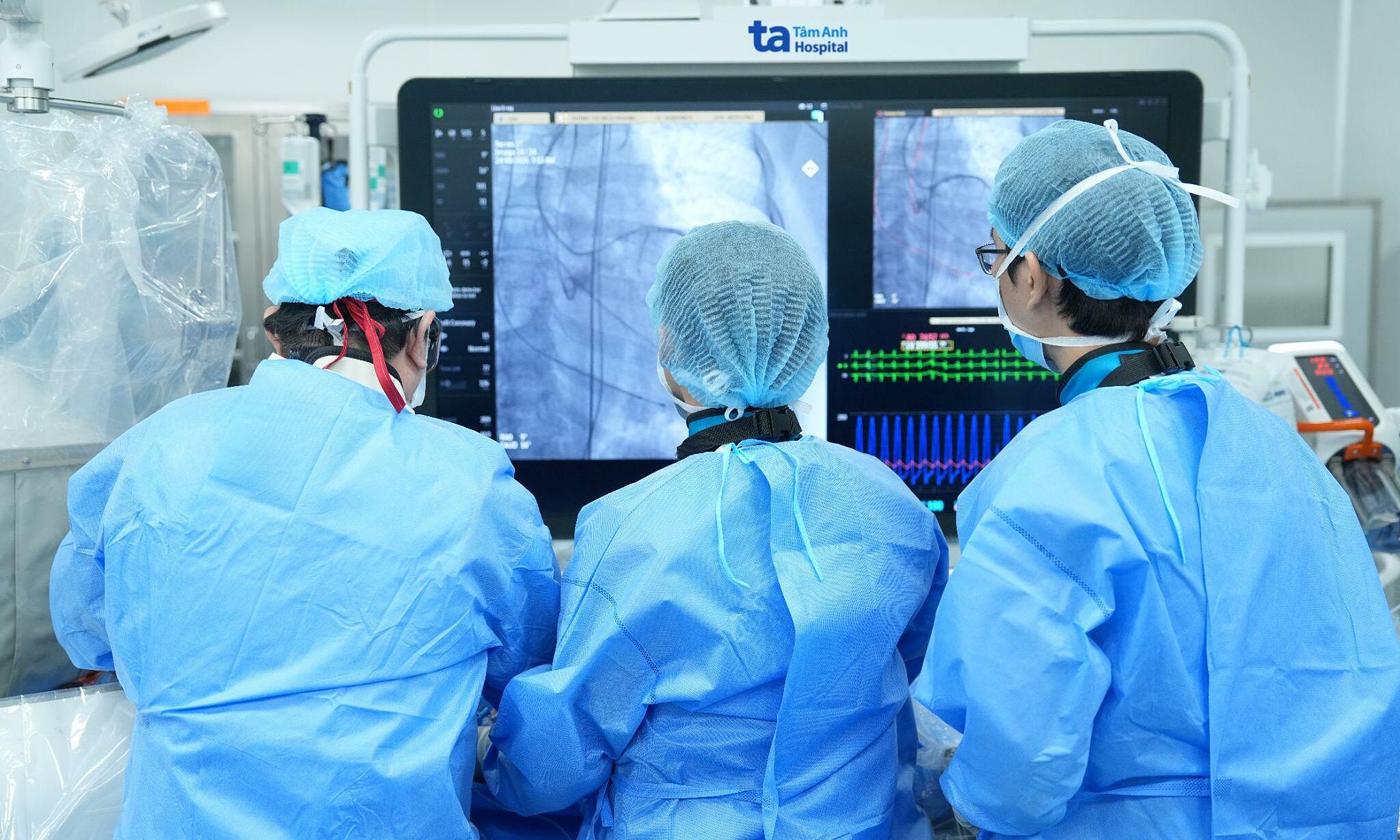
Doctor CKI Nguyen Trung Duc of the Endovascular Intervention Unit at the Interventional Radiology and Imaging Diagnosis Center stated that Mr. Di's hemoptysis could lead to blood entering and obstructing the airways, necessitating immediate bleeding control. Doctors performed digital subtraction angiography (DSA) to examine the vascular system supplying the injured lung area. DSA images revealed numerous proliferating bronchial artery branches concentrated in the lesion in the right upper lung lobe. These abnormal vessels received blood from multiple right broncho-intercostal artery branches and the common bronchial artery trunk.
The team identified the specific vessels requiring intervention and selectively guided a microcatheter deep into each proliferating branch. Once the catheter tip reached the correct position, doctors injected embolic material to selectively occlude the abnormal vessels.
The bleeding source was controlled using embolization, avoiding the need for open-chest surgery. Following the intervention, Mr. Di's hemoptysis decreased, and subsequent specialized tests confirmed pulmonary tuberculosis.
According to Doctor Duc, prolonged tuberculosis lesions can alter the vascular system in the affected lung region. Bronchial arteries proliferate, dilate, and become vulnerable, potentially causing bleeding into the airways, manifested as hemoptysis. Therefore, Mr. Di is receiving pulmonary tuberculosis treatment according to protocol, while continuing to monitor his hemoptysis and lung lesions. After treatment, his symptoms gradually improved, and he attends follow-up appointments as scheduled.
Hemoptysis can stem from various causes, including respiratory infections, prolonged lung damage leading to vascular abnormalities, and pulmonary tuberculosis. Individuals experiencing hemoptysis should seek early medical attention to identify the cause and receive timely treatment.
Nhu Ngoc
*The patient's name has been changed