Doctor Bui Viet Hung, Head of the Vitreoretinal Department at Tam Anh Ha Noi General Hospital’s High-Tech Eye Center, noted the patient's right eye showed grade two proptosis, swelling and pain around the eye, and blurry vision. An MRI of the brain and eye socket revealed a right frontal sinus mucocele measuring 30x51x45 mm. The cyst had eroded the frontal bone, extended into the orbital roof, and pushed against the eyeball, superior rectus muscle, and dura mater. This pressure also compressed the upper retina, causing macular folds and affecting central vision.
Ngoc Duy suffered from amblyopia in his left eye since childhood, while his right eye maintained good vision. Therefore, Doctor Hung prioritized decompressing the optic structures and protecting them to minimize the risk of vision loss.
 |
Image of a large tumor compressing the eyeball. Photo: Tam Anh General Hospital |
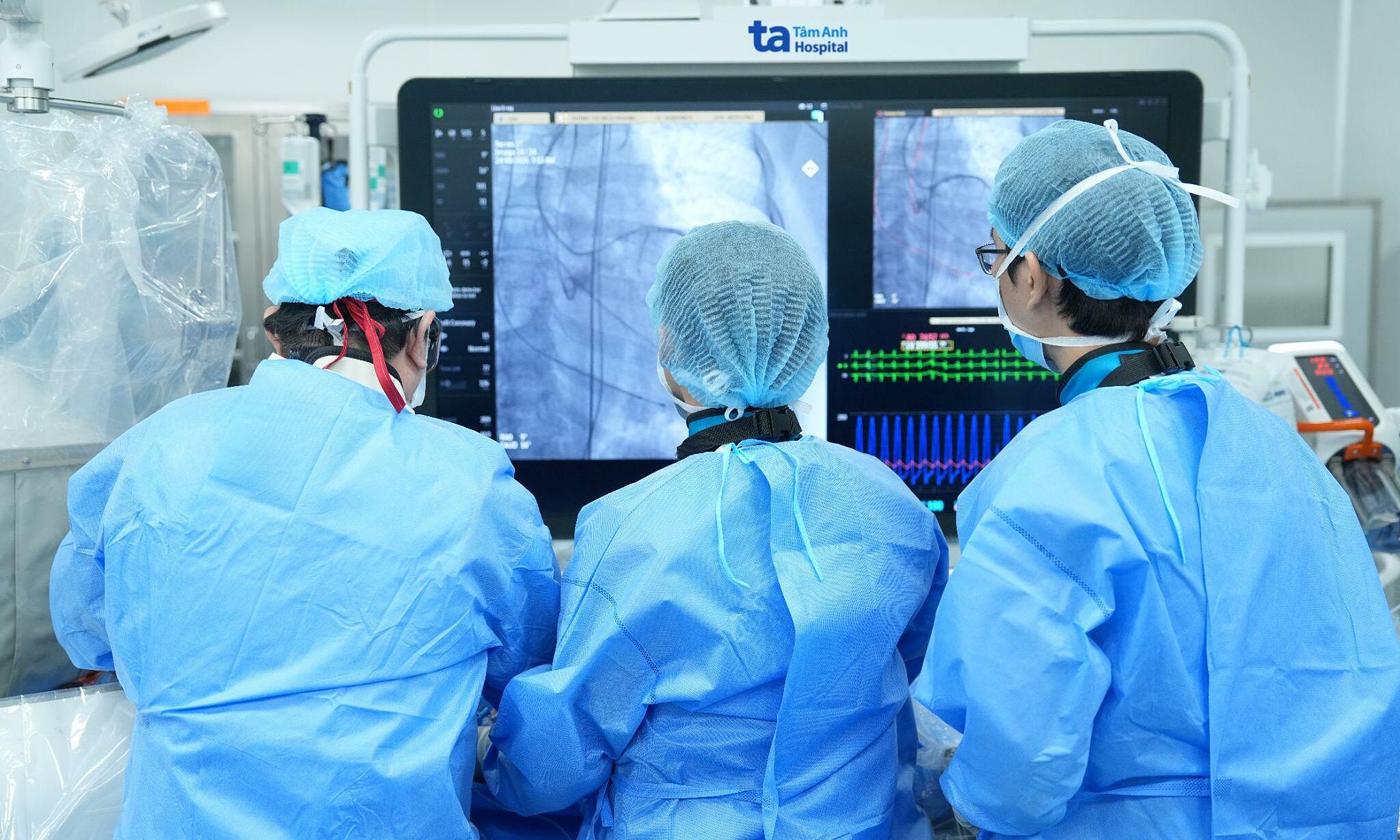
Ngoc Duy has suffered from heart failure since 2016. A recent echocardiogram showed a dilated left heart chamber and a cardiac ejection fraction of only 24% (normal is 50-70%). To prepare for surgery, he underwent a multi-slice coronary computed tomography angiogram and wore a Holter ECG to assess his cardiovascular condition. The High-Tech Eye Center team consulted with specialists from Neurosurgery, Cardiology, and Anesthesiology for surgery to remove the frontal sinus mucocele, decompress the area, and preserve vision in the right eye.
Master of Science, Doctor Le Van Ngan, from the Neurosurgery Department, described the surgery as challenging due to the large lesion close to the skull base, compressing the eye socket, while the patient had severe heart failure, which could cause hemodynamic instability during anesthesia. The anesthesia team performed endotracheal intubation and monitored invasive arterial blood pressure to closely control circulation during the operation.
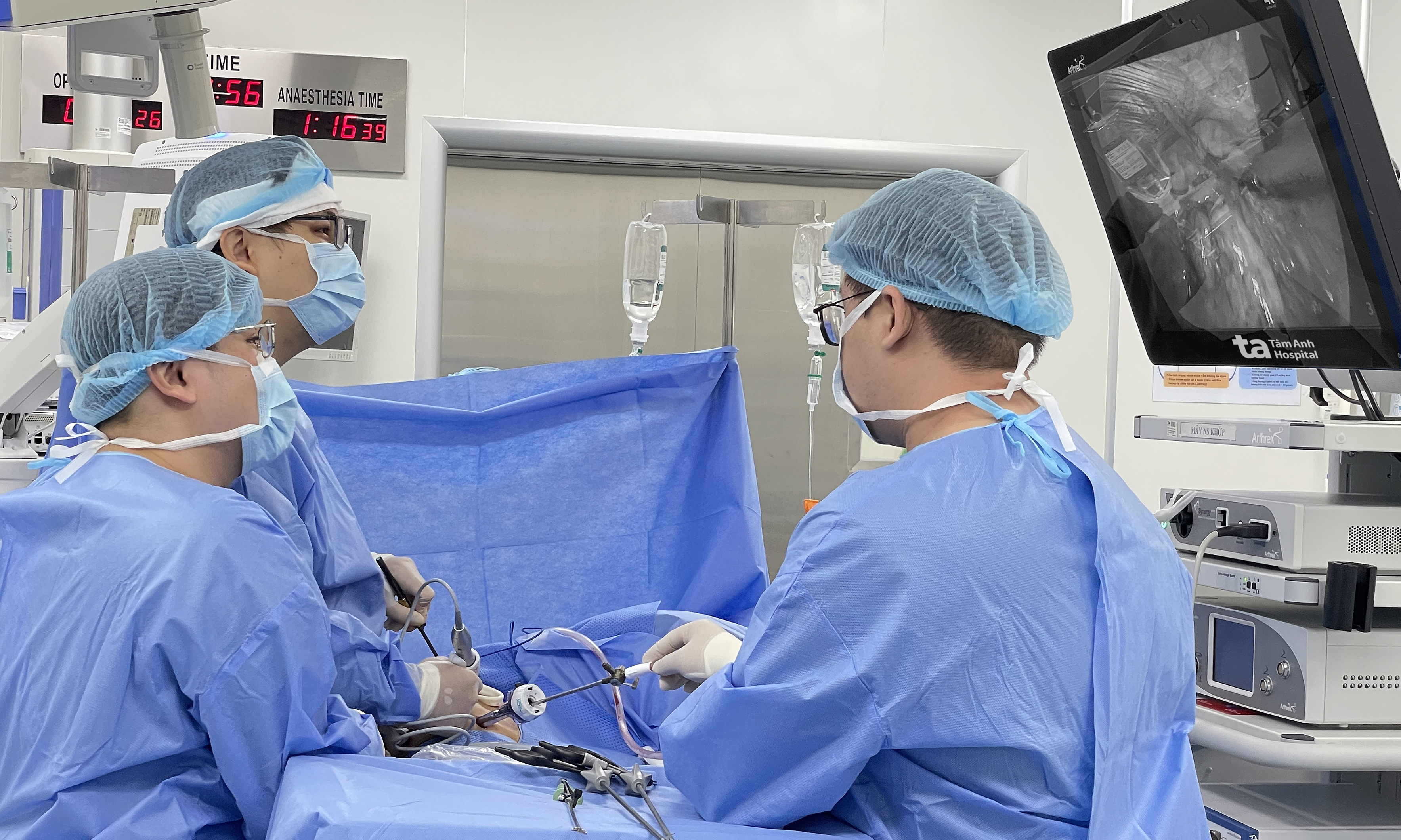
Upon accessing the right frontal area, doctors observed that the frontal bone was eroded and perforated in several places. They aspirated approximately 20 ml of greenish-brown pus for testing. The team used a surgical microscope to completely dissect the cyst lining close to the dura mater, cleaned the frontal sinus, sealed the communication pathway with bone wax and bio-glue, and then reconstructed the bone defect with a titanium patch secured by screws.
 |
The surgical team treats the frontal sinus tumor. Photo: Tam Anh General Hospital |
Pathology confirmed a frontal sinus mucocele with no malignant cells. After surgery, the patient's right eye showed reduced swelling and pain, improved blurry vision, and the eyeball was decompressed, preserving his eyesight. The patient continues to receive follow-up care for his eyes and cardiovascular health.
Doctor Ngan stated that while mucoceles are benign lesions, they can become dangerous if they grow large, erode bone, and spread beyond the sinuses. When they approach the eye socket or skull base, these lesions can affect vision or cause intracranial infection complications.
Patients should seek medical attention if they experience persistent proptosis, swelling or pain around the eye, orbital pain, or blurry vision, especially if symptoms do not improve after initial treatment. Early detection helps manage the lesion before it compresses important structures, protecting vision and reducing the risk of complications.
Thu Giang
| Readers can send ophthalmology questions here for doctors to answer |