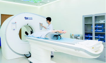
An 8 mm nodular lesion was located in the extraconal space, deep within Hien’s left eye orbit, according to a 3 Tesla MRI scan at Tam Anh General Hospital Ho Chi Minh City.
The orbit is a confined bony cavity containing the eyeball, optic nerve, extraocular muscles, and many important blood vessels. Dr. Mai Hoang Vu, from the Department of Neurosurgery - Spine at the Neuroscience Center, explained that due to the narrow orbital space, even a small tumor like Hien’s can cause a mass effect, compressing the optic nerve, pushing the eyeball forward, and impinging on the extraocular muscles, leading to a gritty, obstructed, and uncomfortable sensation.
While likely a benign orbital tumor, prolonged compression without surgery would affect visual function, Dr. Vu noted.
During a consultation, doctors determined the tumor was located deep behind the eyeball. Traditional approaches, such as accessing from outside the orbit or performing an osteotomy, would involve a longer, more invasive path, risking damage to healthy tissue and prolonging recovery. After careful consideration, the surgical team opted for a direct trans-ocular approach, combining endoscopy with an AI-integrated neuro-navigation system. The goal was to reach the tumor via the shortest possible route, enhancing precision, minimizing trauma, and preserving Hien’s vision. This innovative surgical method was researched and implemented for the first time at Tam Anh Hospital.
Before the surgery, all MRI data of the tumor and surrounding vital structures were input into the neuro-navigation system to create a 3D spatial map. This allowed surgeons to precisely determine the coordinates, approach direction, and safety margins throughout the procedure.
After setting up the navigation system, the team created an entry point through the inner corner of the eye, retracting the upper and lower eyelids to create a working corridor. Through this small incision, the eyeball was gently moved aside instead of being removed, as in previous techniques. Surgeons then created an access path through the sclera – the white protective layer of the eyeball – and introduced a high-resolution endoscope along with microsurgical instruments to reach the area behind the eyeball.
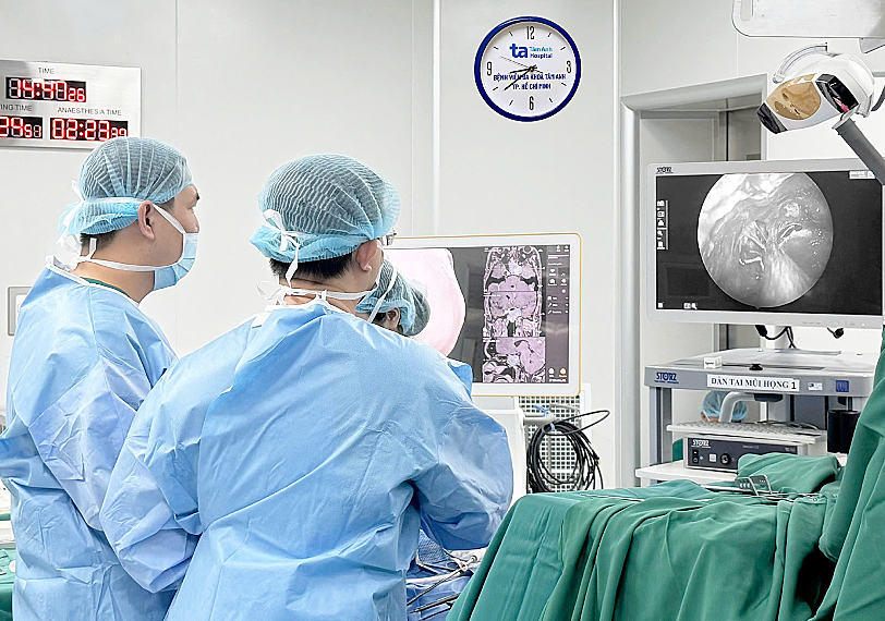 |
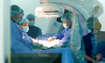
Doctors performing surgery on Hien. Photo: Tam Anh General Hospital |
The team meticulously dissected the tumor in sections, actively controlled bleeding, and completely removed the mass before closing the surgical site with microsutures. A post-operative brain CT scan confirmed the tumor's complete removal and resolution of the mass effect in the left orbit. Following surgery, Hien reported an end to the gritty and obstructed eye sensation, significant improvement in her vision, and no neurological complications.
"The success of this surgery confirms the feasibility and safety of the minimally invasive trans-ocular approach", said Dr. Vu. He added that this method offers an additional treatment option for deep retrobulbar lesions, orbital apex tumors, and certain anterior-lateral skull base tumors, which previously often required highly invasive open surgery.
Orbital and retrobulbar tumors often progress silently and can be overlooked with only a clinical examination. Patients experiencing persistent eye pain, proptosis (bulging eye), restricted eye movement, or decreased vision should seek specialized medical care for an MRI or CT scan. Early detection and timely treatment are crucial to prevent long-term vision-threatening complications.
Trong Nghia
*Patient's name has been changed
| Readers can submit questions about neurological diseases here for doctors to answer. |