The woman, in her second pregnancy, underwent an ultrasound at the 22nd week, revealing pulmonary artery valve stenosis and tricuspid regurgitation in the fetus's heart. An amniocentesis for chromosomal analysis showed normal results. Doctors closely monitored the condition, noting a rapid worsening of the fetal heart disease, with progressive right ventricular hypoplasia. Without intervention during the fetal stage, there was an approximately 40% risk of fetal demise, or the child would suffer from severe right ventricular hypoplasia after birth. In such a scenario, a child born with this condition would face numerous complex surgeries, potentially requiring a heart transplant.
A team of specialists from Tu Du Hospital and Children's Hospital 1, including obstetricians, fetal interventionists, and interventional cardiologists, convened to determine the best course of action. They agreed that fetal heart catheterization was immediately necessary. The objective of this intervention was to improve blood flow through the fetal pulmonary artery valve, allowing the right ventricle to continue developing within the uterus.
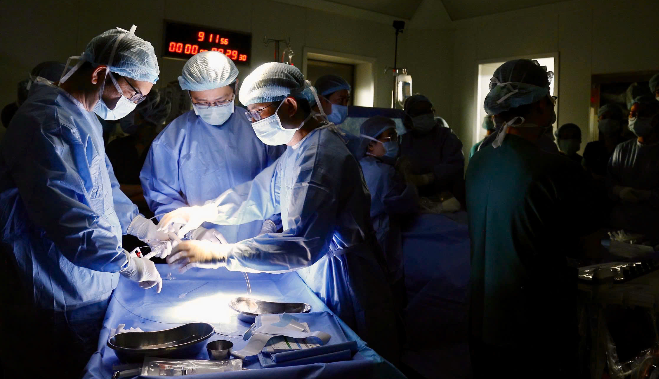 |
The medical team performing the fetal heart intervention on 30/12. Photo: Hospital provided
The pregnant woman received spinal anesthesia, while the fetus was directly anesthetized with specialized medication. Guided by ultrasound, the obstetrics team accessed the right ventricle of the fetal heart through the mother's abdominal wall and uterine cavity. They then performed pulmonary artery valvuloplasty, a critical step in fetal heart catheterization.
At the time of the intervention on the morning of 30/12, the fetus was in a cephalic presentation, tilted to the left, with the placenta located anteriorly. These factors increased the procedure's difficulty. Notably, the fetal arm was draped across its chest, obscuring the path to the heart chambers. The team performed minor adjustments to the fetal position, ensuring safe access to the intervention site, and successfully dilated the pulmonary artery valve on the first needle puncture. Post-intervention checks showed significant improvement in blood flow through the fetal pulmonary artery valve, minimal pericardial effusion, and a stable fetal heart rate.
This case marks the 12th fetal heart catheterization performed in Ho Chi Minh City and the first to be implemented after the Ministry of Health officially approved the list of techniques for fetal heart intervention in Vietnam. This procedure demands a high level of expertise, close inter-hospital collaboration, and is considered a significant achievement in Ho Chi Minh City's specialized medicine.
 |
The pregnant woman and her husband at Tu Du Hospital after the fetal heart intervention. Photo: Hospital provided
Tang Chi Thuong, Director of the Ho Chi Minh City Department of Health, lauded this success, stating it confirms the professional capability, strong inter-hospital coordination, and mastery of advanced fetal heart intervention techniques by Ho Chi Minh City's medical teams. This achievement contributes to standardizing and advancing specialized fetal medicine in Vietnam, promoting regional and international integration.
In May, a 41-year-old woman from Singapore, experiencing her first pregnancy after more than 10 years of infertility through in vitro fertilization, was referred to Vietnam for her child's heart catheterization—a technique few places in the world perform. The pregnant woman later returned home, giving birth in July. The baby girl only required a minor post-birth procedure, a stark contrast to the previous prognosis of "incurable" if the fetal heart catheterization had not been performed earlier.
Le Phuong