

An ultrasound performed on a 32-year-old pregnant woman revealed that the fetus had multiple vertebrae fused together, forming a single bone mass instead of developing separately and being cushioned by intervertebral discs as normal. Additionally, one lumbar vertebra was improperly formed, appearing wedge-shaped (normally cylindrical).
Doctor Nguyen Hoang Long from the Fetal Medicine Unit at Tam Anh General Clinic, District 7, diagnosed the fetus with fused thoracic and lumbar vertebrae, along with hemivertebrae. These conditions could be isolated defects or linked to genetic disorders such as Klippel-Feil syndrome, Jarcho-Levin syndrome, Alagille syndrome, or MURCS association.
Further examination showed that the two lower poles of the fetal kidneys were fused, forming a U-shape. This is a horseshoe kidney, which may be a morphological manifestation of Turner syndrome (XO), Edwards syndrome (trisomy 18), or caudal regression syndrome (CRS). A horseshoe kidney can also be an isolated defect or, in some cases, accompanied by anomalies of the genitourinary system, brain, or cardiovascular system. The fetus also presented with a single umbilical artery; normally, there are two arteries and one vein.
In-depth genetic analysis identified a duplication of chromosome 2 p25.3 in the fetus. According to Clinvar, a database of human genetic variations maintained by the U.S. National Institutes of Health (NIH), the function of this chromosomal duplication is unclear, and it does not manifest specific phenotypic abnormalities. "Based on medical literature, the duplication of chromosome 2 p25.3 may not be related to the fused vertebrae, hemivertebrae, or horseshoe kidney," Doctor Long stated. However, the child's physical, intellectual, and language development will require monitoring later.
Doctor Long predicted that these morphological anomalies would likely not affect the baby's vital functions after birth. Regarding spinal defects, most cases develop normally. Some children are at risk of scoliosis or spinal deformities. After 30 years old, there is a possibility of spinal canal stenosis progressing, causing spinal cord compression and late neurological symptoms such as limb numbness or muscle weakness, which may require orthopedic surgery.
Most cases of horseshoe kidney rarely severely affect kidney function during infancy and early childhood. As adults, patients are at risk of developing urinary complications, including urinary tract infections, kidney stones, ureteropelvic junction obstruction, and vesicoureteral reflux, necessitating regular health check-ups.
Doctors performed regular specialized morphological and fetal echocardiography throughout the pregnancy to closely monitor fetal development. They noted that the spine remained undeformed, the fetus moved normally, and no other abnormalities were detected. At 39 weeks of gestation, a baby girl was born, weighing 2,9 kg, with stable health upon examination. However, the baby requires regular follow-up at each developmental milestone to assess growth, urinary, neurological, and motor functions for early detection of any potential abnormalities.
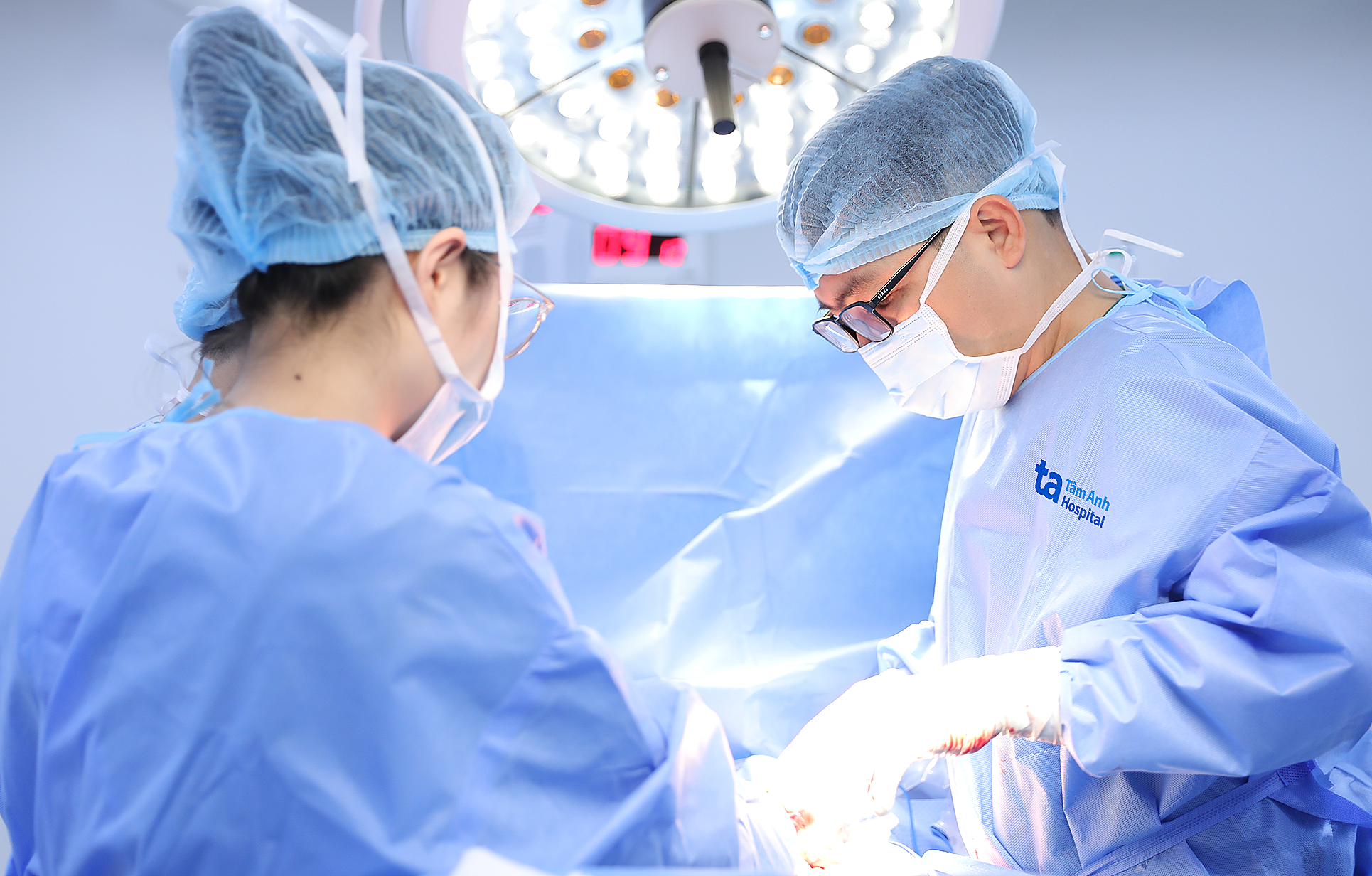 |
The medical team performed a C-section for the pregnant woman. Photo: Tam Anh General Hospital
Doctor Long explained that the impact of anomalies during pregnancy varies depending on the cause, type of defect, and related functions. Some defects can be corrected during pregnancy or treated after birth, ensuring a good quality of life for the child. Pregnant women should undergo regular prenatal check-ups at specialized units to detect any abnormalities early and determine appropriate management.
Ngoc Chau
| Readers can submit questions about obstetrics and gynecology here for doctors to answer. |


