A baby boy, diagnosed with congenital duodenal stenosis during pregnancy, was safely delivered at week 39 at Tam Anh General Hospital, TP HCM. Following birth, the infant underwent successful surgery to correct the condition, creating a clear passage from the stomach through the duodenum to the small intestine. The child is now in stable health. Congenital duodenal stenosis is a partial narrowing of the duodenum, which leads to incomplete intestinal obstruction, preventing food and digestive fluids from flowing properly from the stomach to the intestines.
The condition was first detected at week 22 of pregnancy, when ultrasound revealed duodenal stenosis and subsequent stomach dilation. Doctor Le Quang Hung, from the Fetal Medicine Center at Tam Anh General Hospital, TP HCM, noted that congenital duodenal stenosis affects approximately one in 10,000 newborns. It can be an isolated defect or associated with genetic abnormalities such as Down syndrome (trisomy 21) or Edwards syndrome (trisomy 18). Some cases also involve other malformations affecting the: heart, kidneys, limbs, spine, bones, esophagus, intestines, bile duct, and pancreas. For the fetus, risks include polyhydramnios, preterm birth, stillbirth, and fetal demise. After birth, affected infants may experience vomiting due to milk intolerance, aspiration pneumonia, and electrolyte imbalance.
To assess the prognosis, the expectant mother was advised to undergo amniocentesis, which revealed no genetic abnormalities. Doctor Hung evaluated the case as having a positive prognosis, emphasizing the need for close monitoring throughout the pregnancy to identify any other potential abnormalities. By week 30, the expectant mother developed polyhydramnios because the fetus could not effectively swallow and digest amniotic fluid. This excess amniotic fluid can overdistend the uterus, increasing the risk of early labor and preterm birth. The medical team decided to administer a full course of lung maturation injections to reduce the risk of respiratory distress for the baby, should a preterm birth occur. The mother was also guided on appropriate diet, exercise, and how to count fetal movements at home.
At week 39, ultrasound results showed an amniotic fluid index (AFI) of 31 cm and a largest amniotic pocket of 8,8 cm, both exceeding normal thresholds. This indicated a risk of sudden membrane rupture during labor, coupled with an unstable fetal heart rate. Given these concerns, doctors opted for a C-section. The baby boy was delivered safely, weighing 2,8 kg, and showed no additional abnormal symptoms immediately after birth.
 |
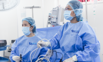
The obstetrics team performing a C-section for the expectant mother. Photo: Tam Anh General Hospital |
The obstetrics team performing a C-section for the expectant mother. Photo: Tam Anh General Hospital
In cases of congenital duodenal atresia, which is a complete obstruction, infants may experience vomiting within the first few hours after birth, especially following their first feeding. The vomit often contains bile, appearing green or yellow. Other accompanying signs include upper abdominal distension, constipation (absence of meconium), jaundice, and low birth weight.
For partial duodenal stenosis, symptoms may manifest later, presenting as abdominal pain, indigestion, malabsorption, or a palpable abdominal mass resulting from localized gastric or duodenal dilation.
Early prenatal detection of duodenal stenosis is crucial for optimal pregnancy management and preparing a comprehensive postnatal care plan. This plan includes interventions such as placing a gastric tube to decompress the stomach, preventing aspiration, and providing intravenous feeding. Doctor Hung recommends that expectant mothers undergo regular prenatal check-ups at specialized medical centers. Such routine visits enable early detection of any abnormalities, ensuring optimal monitoring and treatment to safeguard the health of both mother and baby.
Ngoc Chau
| Readers can submit questions about obstetrics and gynecology here for doctors to answer. |