Dr. Ha To Nguyen, Director of the Fetal Medicine Center at Tam Anh General Hospital, TP HCM, reported that the fetus was diagnosed with cardiac rhabdomyomas. Cardiac rhabdomyomas can be an isolated abnormality or, in 80-90% of cases, linked to Tuberous Sclerosis Complex (TSC). This is an autosomal dominant genetic syndrome, affecting about one in 6,000 children. 30% of cases are inherited, while 70% result from new mutations in the TSC1 gene on chromosome 9 or the TSC2 gene on chromosome 16. Tuberous Sclerosis Complex typically affects the central nervous system, heart, eyes, kidneys, and skin.
According to Dr. Nguyen, heart tumors are often diagnosed during a 20-week morphological ultrasound, commonly found in the ventricular muscle or interventricular septum. If a tumor is large or in a critical location, it can cause arrhythmia, obstruction of blood flow from the heart chambers, heart dysfunction, hydrops fetalis, or stillbirth.
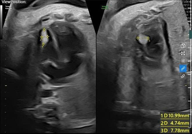 |
A 32-week fetal ultrasound showing a tumor in the left ventricular muscle near the apex. Photo: Tam Anh General Hospital |
After the abnormality was detected, doctors recommended amniocentesis and fetal Magnetic Resonance Imaging (MRI) to further assess for brain lesions and determine if the cardiac rhabdomyoma was isolated or due to a gene mutation related to Tuberous Sclerosis Complex. However, Nguyet and her husband declined the procedures, concerned that invasive intervention might affect the fetus.
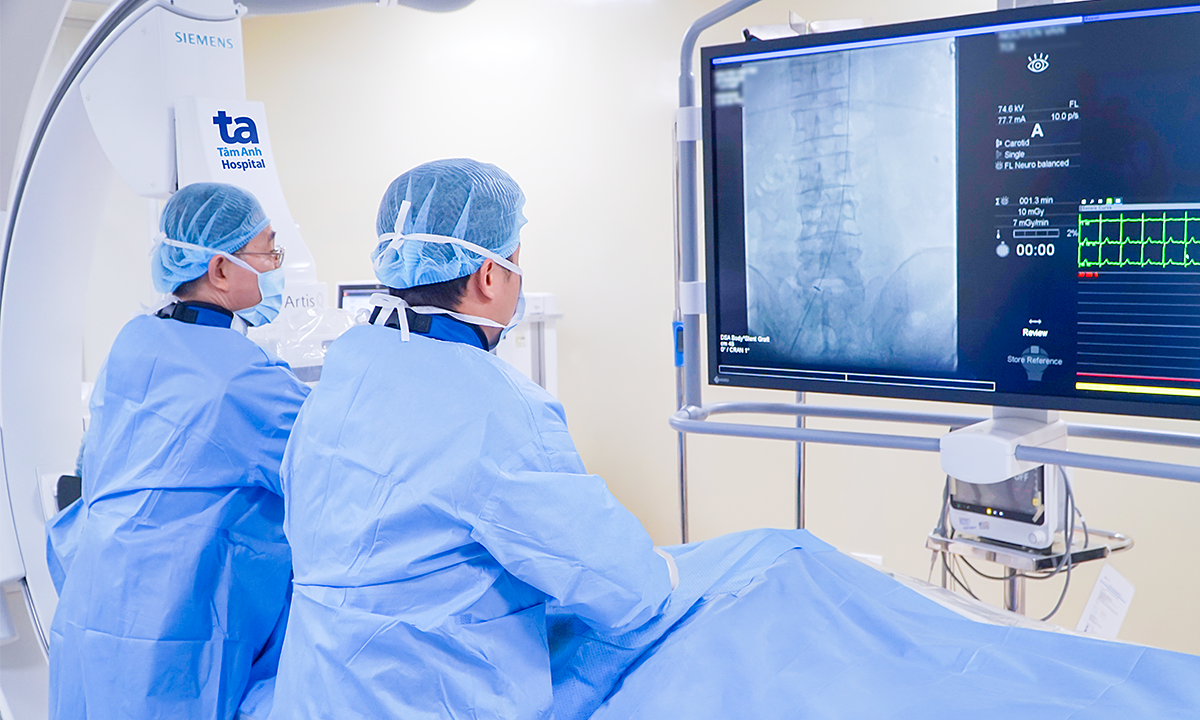
Doctors from the Fetal Medicine and Obstetrics departments, in coordination with cardiologists, closely monitored the fetal heart morphology throughout the pregnancy to control the risk of rapid tumor growth. The tumors increased in size during gestation but did not cause fetal heart failure, the heart rate remained stable, and the fetus developed normally.
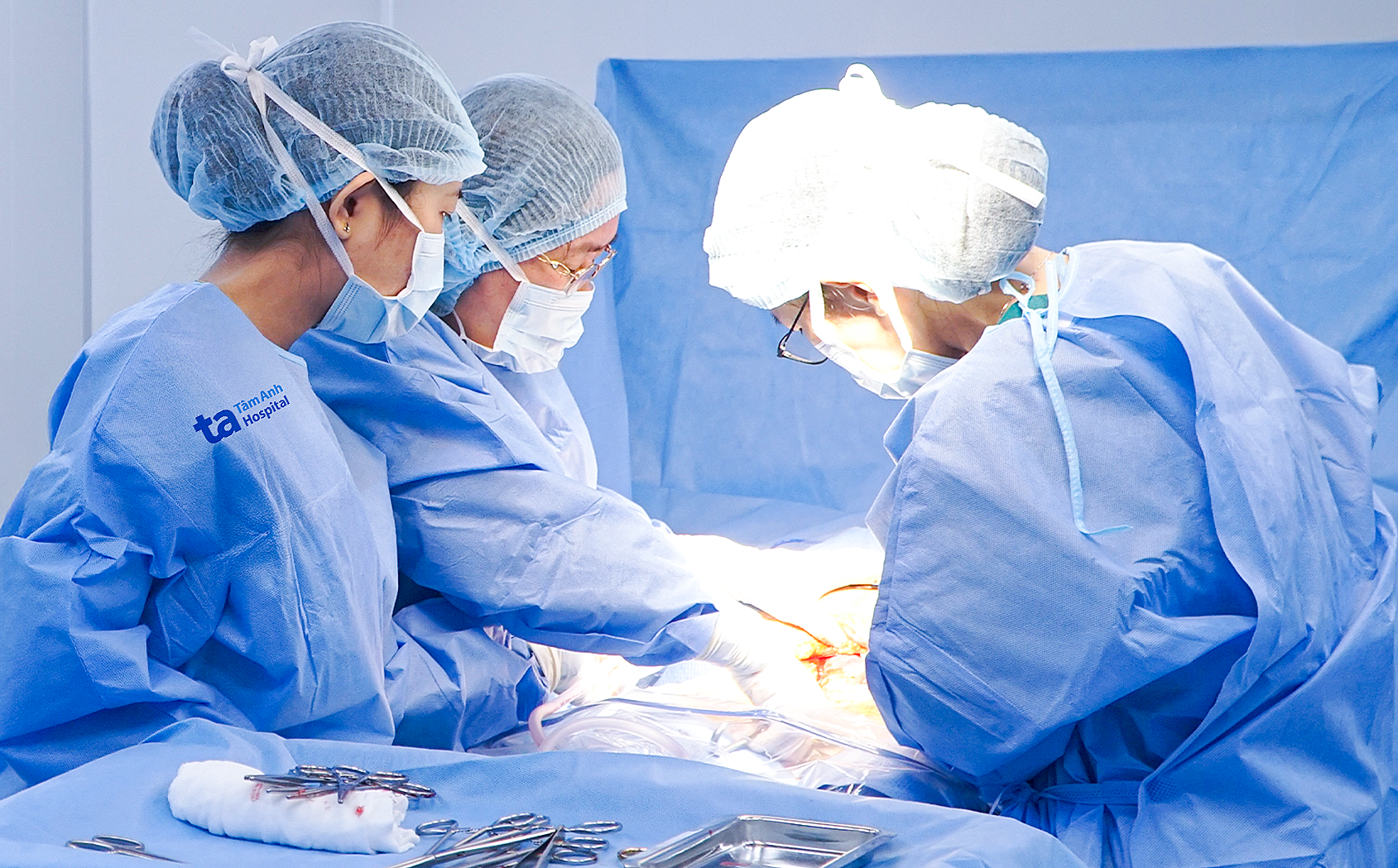 |
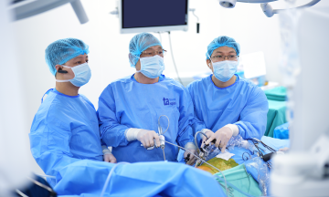
Master of Science, First Degree Specialist Doctor Tran Lam Khoa (right) and his team perform a C-section for Nguyet. Photo: Tam Anh General Hospital |
By the 38th week, a fetal echocardiogram showed that the tumor near the heart's apex did not obstruct the outflow tract or affect the heart valves. Nguyet underwent a C-section, and a healthy baby boy weighing 3,4 kg was safely delivered. A subsequent echocardiogram revealed a 10,5 x 5 mm heart tumor attached to the interventricular septum near the left ventricular apex, and another tumor measuring 2,9 x 4,1 mm near the right ventricle. Currently, the baby's heart function is good.
Dr. Van Thi Thu Huong, from the Cardiology Center, stated that during the fetal stage, cardiac rhabdomyomas tend to grow rapidly in size during the second trimester, influenced by maternal hormones. This rapid growth mostly ceases by the 32nd week. After this point, the tumors typically do not change in size.
According to Dr. Huong, cardiac rhabdomyomas usually appear in the two ventricles, with an equal proportion in the left and right ventricles. Tumor sizes vary, ranging from small (5 mm to 10 mm) to larger than 40 mm.
Most cardiac rhabdomyomas are asymptomatic and are often incidentally discovered during an echocardiogram. However, a significant percentage (15%) experience cardiovascular complications such as heart chamber obstruction, arrhythmia, heart failure, or even death. These symptoms occur with larger tumors or those located near the heart's conduction and pacing system.
In addition to cardiovascular complications, cardiac rhabdomyomas are closely linked to Tuberous Sclerosis Complex, a genetic condition characterized by the formation of benign tumors in multiple organs, particularly the heart and nervous system. Nervous system lesions play a crucial role in the long-term prognosis. Children are at high risk of complications such as seizures, refractory epilepsy, psychomotor developmental delay, and autism spectrum disorder. Structural neurological lesions in Tuberous Sclerosis Complex include cortical tubers, subependymal nodules, giant cell astrocytomas, and white matter lesions. These lesions can appear early in the fetal stage, while clinical manifestations often emerge later after birth.
Therefore, a comprehensive evaluation of the clinical and imaging characteristics of cardiovascular and neurological lesions in children with cardiac rhabdomyomas is crucial. This helps in early detection of complications and allows for appropriate treatment and preventive measures, improving patient prognosis.
Experts recommend that pregnant women undergo all scheduled ultrasounds, especially in the second and third trimesters, to detect fetal heart abnormalities and other congenital defects early. Early detection of abnormalities during pregnancy enables doctors to develop a monitoring plan, coordinate multidisciplinary care prenatally, prepare for a safe delivery, and intervene for the baby immediately after birth if necessary.
Tue Diem
* Character names have been changed
| Readers can submit questions about obstetrics and gynecology here for doctors to answer |