Frank, 67, discovered a kidney tumor during a routine check-up in the US last month, despite no back pain or blood in his urine. He and his wife traveled to Vietnam for treatment at Tam Anh General Hospital, Ho Chi Minh City.
A CT scan revealed a left kidney tumor measuring approximately 3,2 x 3,5 cm. Master, Doctor Nguyen Tan Cuong, Head of Urology B and Head of the Urological Oncology Unit at the Center for Urology - Nephrology - Andrology, suspected renal cell carcinoma (RCC). The tumor was on the posterior side of the renal hilum, deep within the renal sinus—an area with major blood vessels and the renal calyces and pelvis system—presenting a difficult surgical challenge.
Frank's medical history included hypertension, type 2 diabetes, and coronary artery disease. He had previously undergone coronary stent placement and coronary artery bypass surgery about one year ago, increasing his surgical risk. Doctor Cuong explained that careless intervention could damage renal blood vessels, leading to severe bleeding or injury to the renal calyces and pelvis system, causing urine leakage post-surgery.
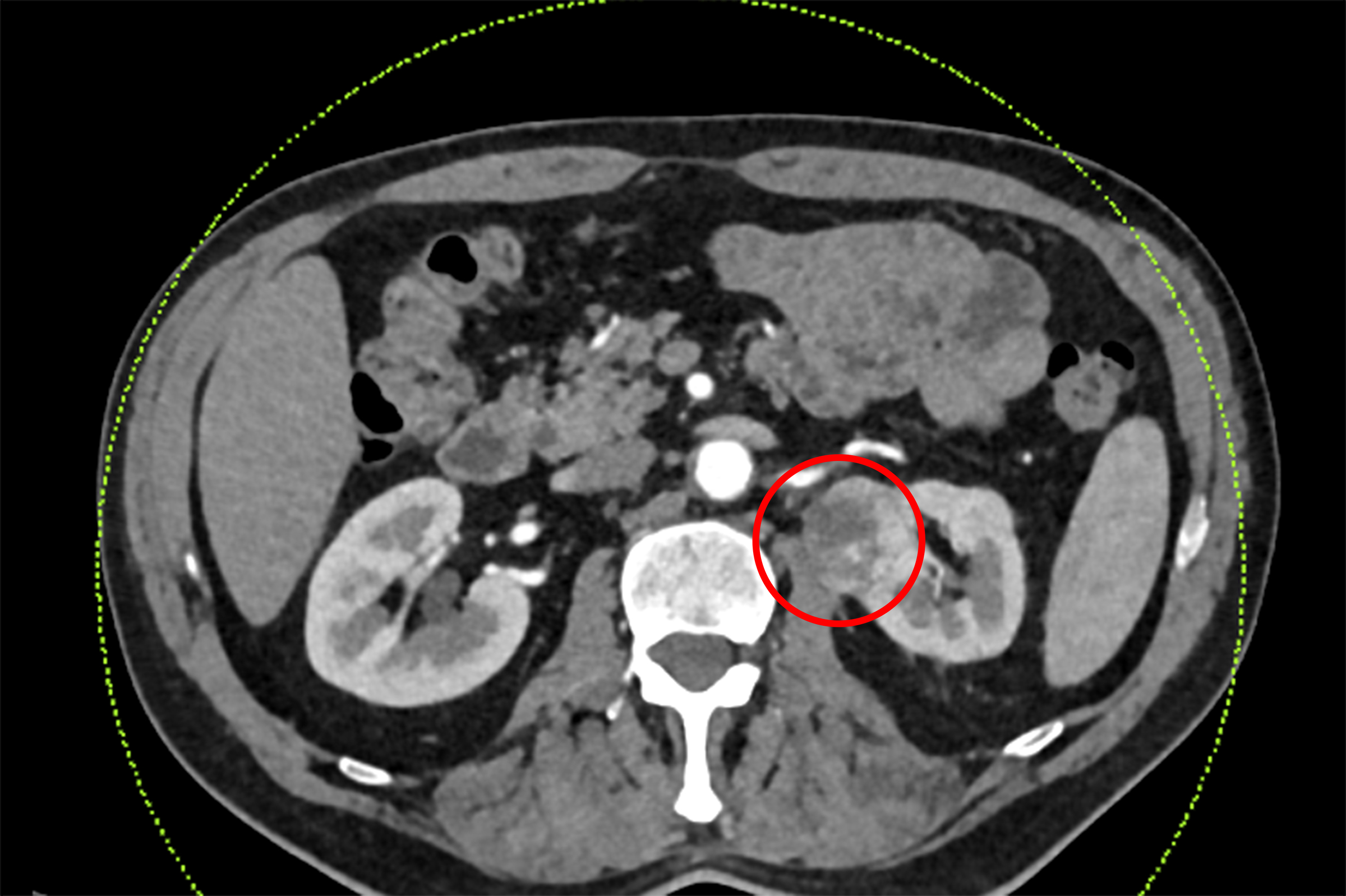 |
The patient's suspected cancerous left kidney tumor on a CT scan. *Photo: Tam Anh General Hospital* |
A multidisciplinary team of urologists, cardiologists, and anesthesiologists consulted, assessing a 6% risk of stroke before, during, and after surgery. The team decided to temporarily stop antiplatelet medication five days before the procedure and resume it early post-operation, balancing the risks of bleeding and thrombosis.
Frank underwent laparoscopic surgery with the new-generation Da Vinci Xi robot system. The goal was to remove the entire tumor while preserving healthy kidney tissue. During the procedure, Doctor Cuong and his team, Master, Doctor Nguyen Truong Hoan and Doctor, Level I Specialist Phan Truong Nam, observed that the tumor's actual position was more complex than external imaging suggested. From the front, through the laparoscopic route in the abdominal cavity, they could barely see the entire tumor. The team meticulously dissected the renal hilum, approaching from front to back to precisely identify the lesion's location and boundaries, while controlling blood vessel structures before resection.
The high-resolution 3D camera system magnified the surgical field 10-15 times, helping the team visualize deep, narrow anatomical structures. Flexible robotic arms enabled doctors to precisely dissect the tumor, suture, and cut in confined spaces.
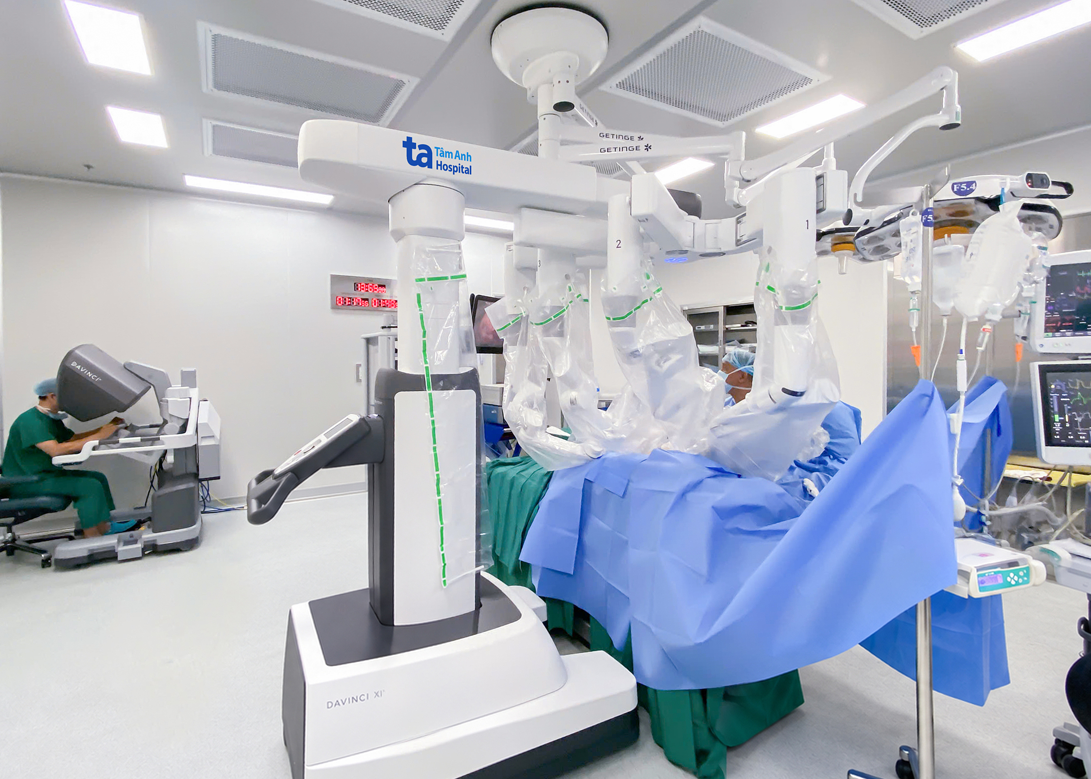 |
Doctor Cuong operating the Da Vinci Xi robot to remove Frank's left kidney tumor. *Photo: Tam Anh General Hospital* |
After three hours, the tumor was completely removed, and healthy kidney tissue was preserved. Subsequent histopathology confirmed the tumor as renal cell carcinoma.
A few hours post-surgery, Frank could eat, walk around his room, and his health was stable. He noted that the examination, testing, and treatment process in Vietnam was efficient, and the cost was one-tenth of what he expected in the US. He received his medical records, surgical report, and histopathology results in English to facilitate follow-up with his doctors in the US.
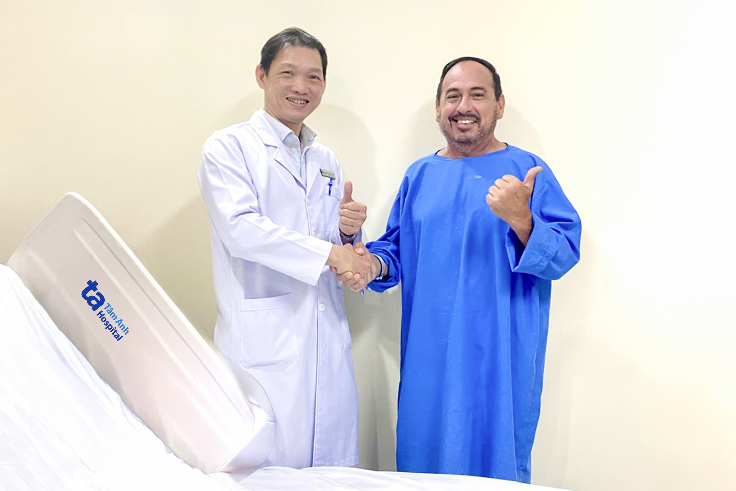 |
Frank (right) could walk just a few hours after robotic left kidney tumor surgery. *Photo: Tam Anh General Hospital* |
According to Doctor Cuong, renal cell carcinoma is the most common malignant kidney cancer in adults, accounting for 80-90% of kidney cancer cases. In its early stages, the disease often presents no clear symptoms and may be incidentally discovered via ultrasound, CT, or MRI.
Not all kidney tumors require immediate surgery. For small tumors, especially in elderly patients or those with high-risk underlying conditions, doctors may consider imaging surveillance or other treatment methods. When the tumor is localized and can be safely operated on, doctors prioritize partial nephrectomy. Total nephrectomy may be indicated when the tumor is large, extensively invasive, or its location and characteristics are unsuitable for kidney preservation.
Doctor Cuong recommends regular health check-ups every 6-12 months, especially for those with risk factors, to detect abnormalities early and ensure timely treatment.
Ha Thanh
| Readers can ask questions about kidney disease here for doctors to answer |