Both lung X-rays and low-dose lung CT are imaging methods that use X-rays to examine the chest, assisting in the detection and monitoring of respiratory diseases. While both employ X-rays, they differ in their imaging principles, lesion detection capabilities, and clinical indications.
A lung X-ray captures a comprehensive image of the chest on a single plane, allowing doctors to assess the lungs, heart, and pleura. Dr. Nguyen Xuan Phong, from the Department of Diagnostic Imaging at Tam Anh General Hospital, Hanoi, notes that it is often the initial imaging test ordered for various symptoms such as: cough, fever, chest pain, and shortness of breath. This method can identify common conditions such as: pneumonia, pulmonary tuberculosis, pleural effusion, pneumothorax, pulmonary edema, or an enlarged heart. Lung X-rays also help monitor treatment effectiveness for many respiratory patients.
Lung CT scans use X-rays to capture images of the chest from various angles. A computer then processes this data to create multiple thin cross-sectional slices of the lungs. This detailed view allows doctors to identify very small lesions or those obscured by the heart, ribs, or other structures, which are often difficult to see on a standard X-ray.
According to Dr. Phong, low-dose lung CT is optimized to reduce X-ray exposure compared to conventional chest CT. It maintains sufficient image quality to detect small lung nodules and suspicious lesions, making it an effective tool for lung cancer screening.
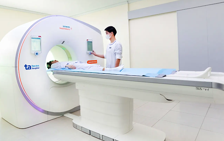 |
Low-dose lung CT screens for lung cancer while reducing X-ray exposure compared to conventional CT. *Photo: Tam Anh General Hospital*
Individuals at high risk for lung cancer include older adults with a long history of heavy smoking (especially those who have smoked one pack per day for 20 years or two packs per day for 10 years), and those with a family history of lung cancer.
Current CT technology has advanced significantly, featuring 32-slice, 64-slice, and 128-slice systems. Some newer generation CT systems can reconstruct over 100,000 slices in a single scan, allowing doctors to observe each region of the lung parenchyma in detail and thus enhancing the detection of very small lesions at an early stage.
Dr. Phong states that for most routine check-ups or common respiratory symptoms, a lung X-ray is typically the first choice. This is due to its ability to detect many common conditions and its quick procedure time. However, if X-ray results are inconclusive, show abnormalities needing further investigation, or if a patient is at high risk for lung cancer, a doctor may then recommend a low-dose lung CT.
Doctors advise against individuals deciding on their own to undergo an X-ray or CT scan during health check-ups or when experiencing respiratory symptoms. After an examination and review of medical history, a doctor will determine the most suitable imaging method based on the patient's symptoms, risk factors, and diagnostic objectives.
Khue Lam
| Readers can submit questions about respiratory diseases here for doctors to answer. |