Ms. Trang, 46, recently underwent a successful embolization procedure at Tam Anh General Hospital Hanoi, significantly reducing the size of multiple liver hemangiomas. She had presented with right upper quadrant pain and bloating, leading to an MRI scan that revealed numerous benign vascular tumors in her liver. The largest of these, located in her right liver near the capsule, measured approximately 86x71x70 mm.
Doctors opted for selective embolization to treat the condition. Following local anesthesia, the medical team accessed Ms. Trang's right femoral artery. A catheter was then guided through the vascular system to the abdominal aorta, celiac artery, and common hepatic artery, with precision guidance from the Artis Pheno robotic system's digital subtraction angiography (DSA) technology.
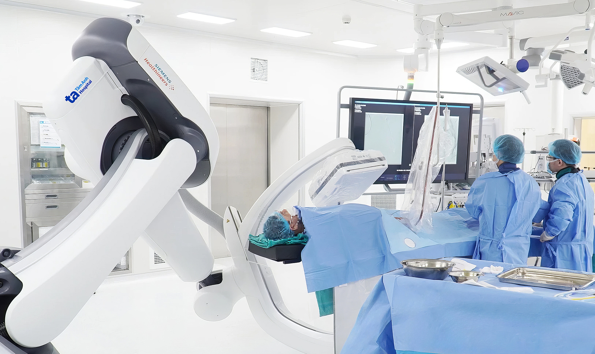 |
The embolization team treating Ms. Trang. Photo: Tam Anh General Hospital |
The procedure involved selectively targeting the arterial branches feeding the hemangiomas and embolizing them with a mixture of oil-soluble contrast agent and a sclerosing agent. Immediately after the intervention, imaging confirmed a 90% reduction in blood flow to the tumors, while preserving healthy liver branches.
One month post-procedure, a follow-up MRI showed a notable decrease in the size of the large tumors in Ms. Trang's right liver. She has since resumed normal activities, and her liver indices remain stable. A follow-up appointment is scheduled for 6 months.
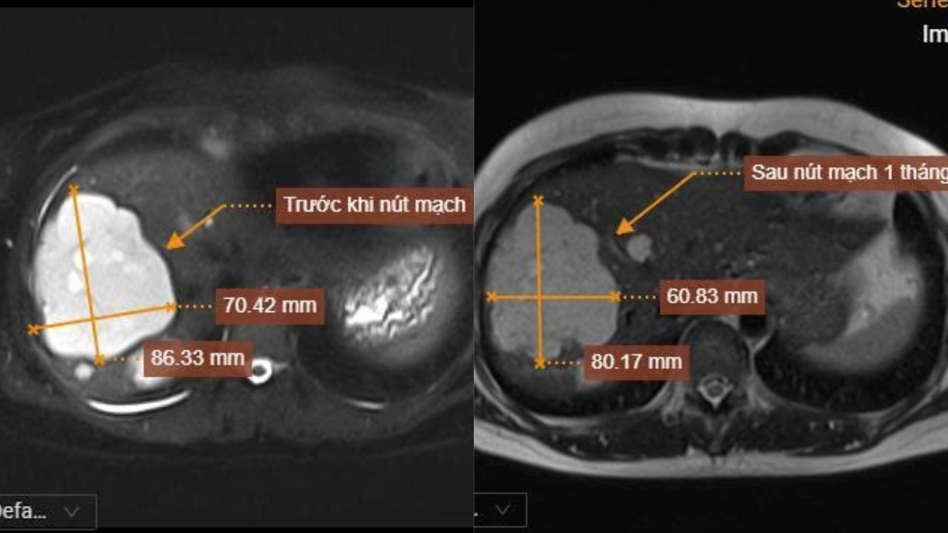 |
An MRI scan shows the largest liver hemangioma in Ms. Trang's liver reduced in size one month after embolization. Photo: Tam Anh General Hospital |
Doctor Nguyen Duy Trinh, Deputy Director of the Center for Diagnostic Imaging and Interventional Radiology, explained that liver hemangiomas are common benign lesions. Typically, small hemangiomas, measuring under 5 cm and located entirely within the liver parenchyma, only require periodic monitoring. However, larger hemangiomas, those that are progressing in size over several months, or those situated in specific locations that could compress veins and bile ducts, carry a risk of rupture and are often candidates for intervention.
According to doctor Trinh, performing embolization on multiple liver hemangiomas during a single intervention helps reduce overall treatment duration and minimizes the number of invasive procedures for the patient. He added that following embolization, tumors typically begin to atrophy within a few months, with the most significant size reduction observed between 3 and 6 months.
Hepatic artery embolization (HAE) works by causing sclerosis of the arterial branches that supply blood to the tumors. This process leads to necrosis and a gradual reduction in the size of the liver hemangioma over time. This minimally invasive technique, guided by AI-integrated angiography machines, ensures high accuracy and effectiveness in treatment.
Hieu Nguyen
*Patient's name has been changed