Dr. Vo Ngoc Bich, from the Department of Hepatobiliary and Pancreatic Surgery, Center for Endoscopy and Gastrointestinal Endoscopic Surgery at Tam Anh General Hospital, TP HCM, stated that Nhan's cystic lesion in the pancreatic head was not a typical pseudocyst often seen after acute pancreatitis. This was because the patient had no history of alcohol consumption, a common risk factor for such conditions.
An MRI scan revealed the cyst was located within the pancreatic head parenchyma, communicating with the main pancreatic duct and accessory pancreatic ducts. Imaging showed irregular dilation of the main pancreatic duct in the body and tail regions, with accessory pancreatic ducts in the pancreatic head also dilated. The cyst likely contained mucinous fluid, accompanied by compression of the distal common bile duct, causing intrahepatic and extrahepatic bile duct dilation. The pancreatic head displayed fibrotic inflammation and adhesive retraction, deforming the pancreatic duct system. Endoscopic ultrasound confirmed the lesion as an intraductal papillary mucinous neoplasm (IPMN) in the pancreatic head.
According to Dr. Bich, intraductal papillary mucinous neoplasm is a type of pre-cancerous lesion. Initially, the tumor may be benign, but it carries a high risk of progressing to cancer, especially when it communicates with the main pancreatic duct, causing duct dilation and exhibiting mural nodules within the cyst, as seen in Nhan's case. The rate of malignant transformation in such cases can reach 60-70%.
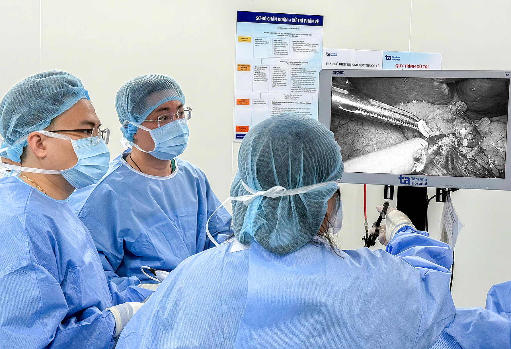 |
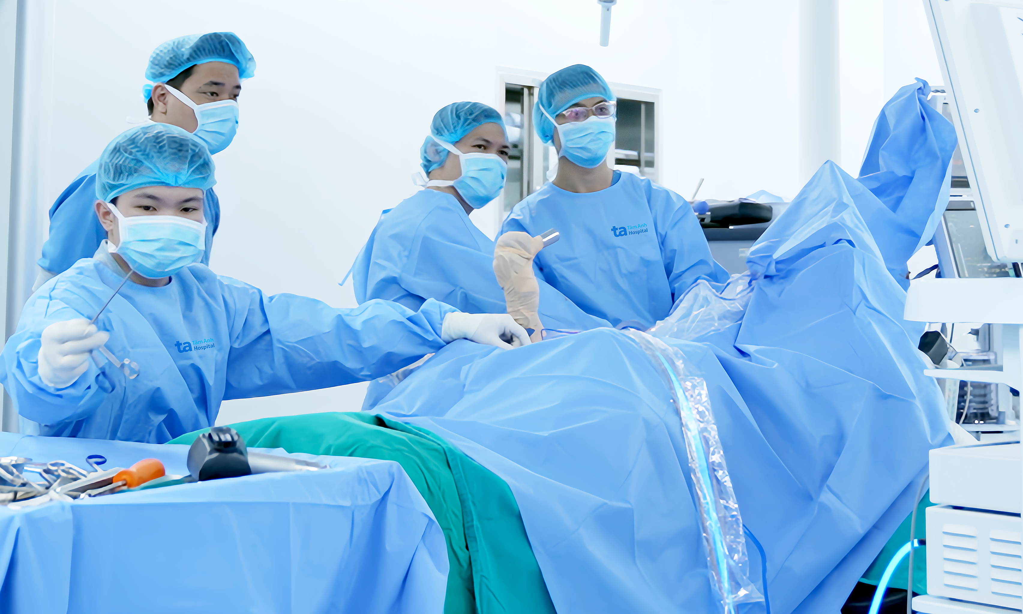
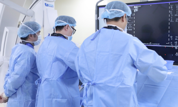
Dr. Bich (right) performs a pancreaticoduodenectomy for Nhan. Photo: Tam Anh General Hospital |
Pancreatic cancer has a poor prognosis and low survival rates due to late detection. For high-risk pre-cancerous lesions, surgery is the only method to completely remove the lesion, reducing the risk of cancer formation and spread.
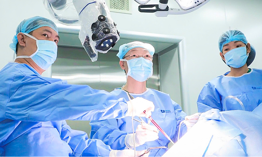
Following a consultation, doctors recommended a pancreaticoduodenectomy (Whipple procedure), which involves removing the pancreatic head, duodenum, a portion of the bile duct, the proximal jejunum, and the lower stomach.
Due to Nhan's history of pancreatitis, the pancreatic head exhibited complex inflammatory adhesions. Surgeons created an additional small access route to dissect and free the adherent tissues, ensuring the safety of critical vascular structures.
After removing the pancreaticoduodenal mass, the surgical team restored digestive continuity by connecting the intestine to the bile duct, the intestine to the pancreatic duct, and performing a gastrojejunostomy, which helped restore the patient's digestive function.
Post-surgery, Nhan recovered well, with no signs of pancreatic fistula – a dangerous complication. He was discharged after 10 days. The patient received guidance on an appropriate diet and will undergo regular follow-up every 6 months for the first two years to detect any abnormalities early.
 |
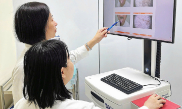
Dr. Bich (left) consults with Nhan during a follow-up visit. Photo: Tam Anh General Hospital |
Doctors advise individuals experiencing persistent abdominal pain, recurrent pancreatitis, or unexplained digestive abnormalities to seek early consultation at specialized medical facilities for accurate diagnosis. Early detection of pre-cancerous lesions like IPMN is crucial for complete treatment and preventing pancreatic cancer, which remains a leading cause of cancer mortality.
Quyen Phan
| Readers can submit questions about digestive diseases here for doctors to answer. |